
|
Figure 5
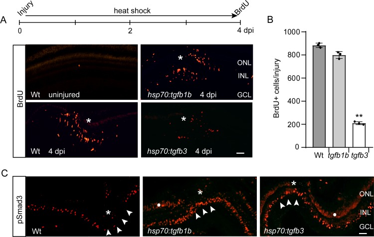
Tgfb1b and Tgfb3 stimulate pSmad3 expression, but only Tgfb3 inhibits injury-dependent MG proliferation.
(A) Top illustration is experimental time line. Bottom panels show BrdU immunofluorescence on retinal sections from uninjured and injured, heat shock-treated Wt, hsp70:tgfb1b, and hsp70:tgfb3 transgenic fish. (B) Quantification of data in (A). (C) pSmad3 immunofluorescence on retinal sections from uninjured and injured, heat shock-treated Wt, hsp70:tgfb1b, and hsp70:tgfb3 transgenic fish. Arrowheads point to the INL at the injury site. Asterisk marks the injury site. White dot in two right-hand panels marks non-specific autofluorescence in the photoreceptor layer. Scale bar is 50 microns. Error bars are SD. **p<0.01.