
Image
|
Figure Caption
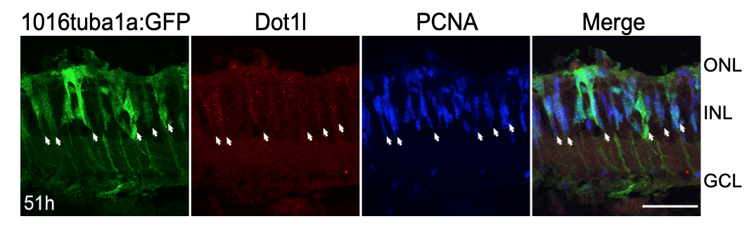
Fig. S1
Dot1l is expressed in proliferating MG after photoreceptor loss. Related to Figure 3. Dot1l (red), GFP, and PCNA (blue) immunostaining after 51 hr intense light lesioning in Tg(1016tuba1a:gfp) retinas. Dot1l is expressed in proliferating MG. Arrows indicate GFP+/PCNA+ dedifferentiated MG that express Dot1l. Scale bar 50um.
Acknowledgments
This image is the copyrighted work of the attributed author or publisher, and
ZFIN has permission only to display this image to its users.
Additional permissions should be obtained from the applicable author or publisher of the image.
Full text @ Cell Rep.