
IMAGE
Fig. S3
- ID
- ZDB-IMAGE-200514-40
- Publication
- Weber et al., 2020 - Zebrafish disease model of human RNASET2 deficient cystic leukoencephalopathy displays abnormalities in early microglia
- All Figures
- Figures for Weber et al., 2020
Image
|
Figure Caption
Fig. S3
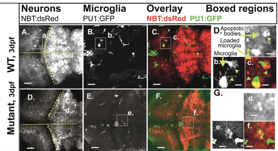
Images of engulfed apoptotic neurons at 3 dpf
Compound transgenic reporter strain zebrafish embryos Tg:(PU1:GFP)/Tg:(NBT:dsRed) were analyzed by in vivo CLSM at 3 dpf. Representative projections of confocal z-stacks through the brain: A, D: NBT:dsRed (neurons); B ,E: PU1:GFP (microglia). C, F: overlays of the respective panels (A,B and C,D). Boxed regions, increased images: a., d.: apoptotic neurons; b., e.: microglia; c. f.: microglia with engulfed apoptotic bodies of neurons (= loaded microglia; arrows). Size markers 40 and 10 μm.
Acknowledgments
This image is the copyrighted work of the attributed author or publisher, and
ZFIN has permission only to display this image to its users.
Additional permissions should be obtained from the applicable author or publisher of the image.
Full text @ Biol. Open