
|
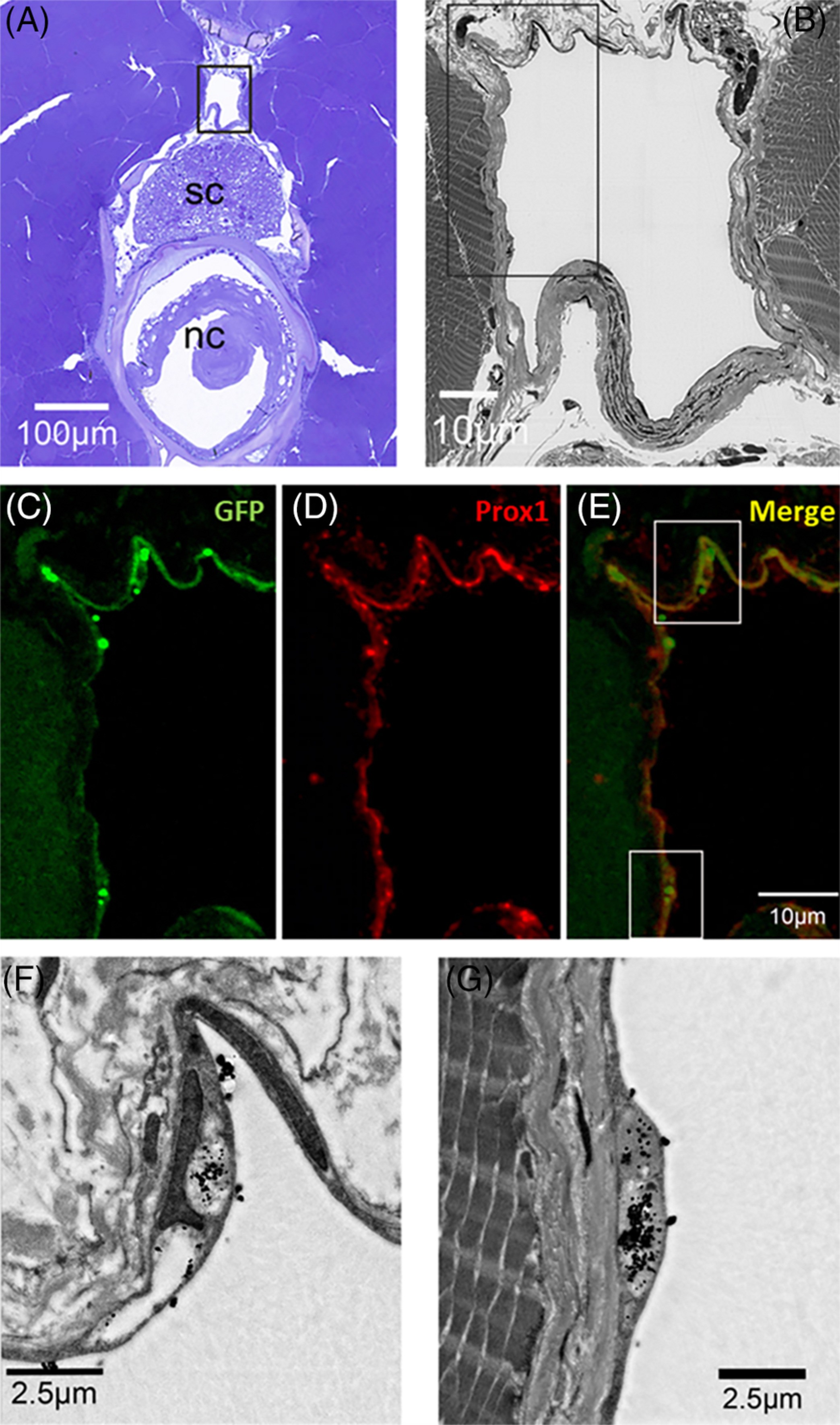
Fig. 4 Correlative scanning microscopy showing cinnabar accumulation in Prox1‐ and EGFP‐immunopositive endothelia of the spinal lymphatic duct in pFLT4‐EGFP adult medaka. A, Cross‐section (toluidine blue) of the trunk to show the relative position of the spinal lymphatic duct (in black frame) in relation to the spinal cord (sc) and notochord (nc). B, Dark‐field SEM image of the spinal lymphatic duct in the frame of Figure 4A. C‐E, Immunostained lymphatic endothelia lining the spinal lymphatic duct in the frame of Figure 4B. C‐E show GFP (green), Prox1 (red), and GFP+ Prox1 (merge) expression, respectively. F,G, Dark‐field SEM images of lymphatic endothelium in the frames of Figure 4E. Note the dense black particles (cinnabar) in/on the endothelium