
|
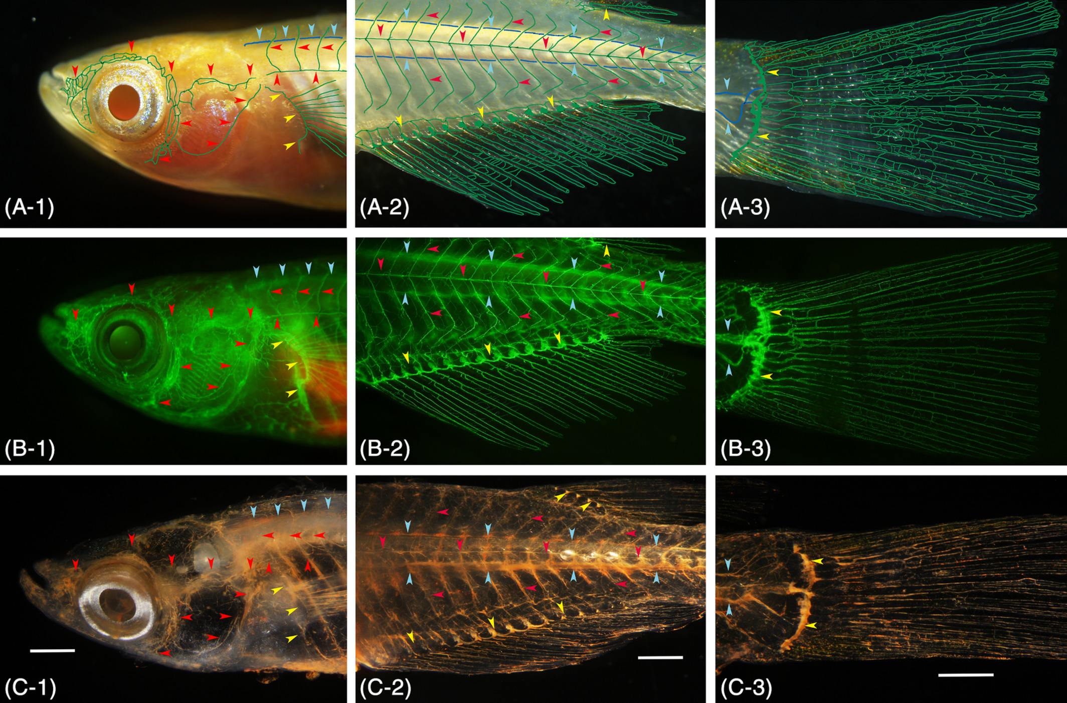
Fig. 2 Gross anatomical identification of the lymphatic vascular system in pFLT4‐EGFP transgenic medaka (adult). A, Conventional stereomicroscopy image of live fish. Superficial (green) and deep (blue) lymphatic ducts and the lymphatic ducts of an appendage (green) are diagrammatically delineated. B, Fluorescence light microscopy image of the same live fish. C, Conventional stereomicroscopy image of the same fish 24 hours after the intraperitoneal cinnabar dye injection. All of the functional lymphatic vascular system was labeled with cinnabar. To verify parietal lymphatic ducts precisely, the brain, gills, and visceral organs were removed from the fixed fish, and the specimen was then cleared with pure glycerol. A‐1, B‐1, C‐1, superficial lymphatic ducts of the cranial region (ie, facial, jugular, opercular, and orbital) and of the trunk (ie, lateral with metameric dorsal and ventral tributaries) are indicated by red arrowheads. Blue and yellow arrowheads indicate the deep lymphatic duct (ie, spinal) and lymphatic vascular system of the pectoral fin on the trunk. A‐2, B‐2, C‐2, the superficial lymphatic system of the trunk and tail (ie, lateral) is indicated by red arrowheads. Blue and yellow arrowheads indicate the deep lymphatic duct (ie, spinal and cardinal‐caudal) and lymphatic vascular systems of the fin (anal and dorsal). A‐3, B‐3, C‐3, lymphatic vascular system of the caudal fin. Blue and yellow arrowheads indicate the deep lymphatic ducts (ie, spinal and caudal) and proximal lymphatic duct of the caudal fin. White bar; 1 mm