
|
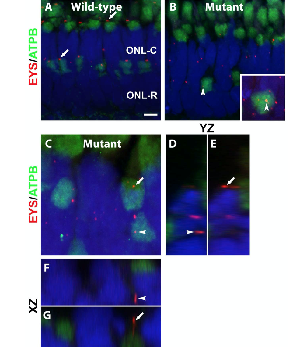
Fig. S1
Mislocalized EYS in pomgnt1 mutant photoreceptors was not localized with mitochondria.
Cryosections of zebrafish eyes were double stained with anti-EYS (red) and with antibody against mitochondrial marker ATPB (green). The sections were counter-stained with DAPI to visualize nuclei (blue).
-
(A) Wild-type. In the wild-type retina, ATPB antibody labeled the ellipsoid region of photoreceptors with EYS immunoreactivity observed on the apical side of ATPB reactivity (arrows).
-
(B) Homozygous pomgnt1sny7 mutant. Most EYS immunoreactive puncta were localized in the outer nuclear layer. Some appeared to be over the ATPB immunoreactive domain (arrowheads)
(C-G) Mutant. Max projection image and its orthogonal views of EYS puncta that appeared to be over the ATPB immunoreactive domains. Of 81 EYS immunoreactive puncta that appeared to overlap with ATPB immunoreactivity, none were within the ATPB immunoreactive domain, indicating that mislocalized EYS were not within mitochondria.
Scale bar in A: = 4.36 m for A-B, 2 m for C.