IMAGE

Fig 4

ID
ZDB-IMAGE-200429-4
Source
Figures for Arribat et al., 2020
Image
Figure Caption

Fig 4 Spastin isoforms in human.

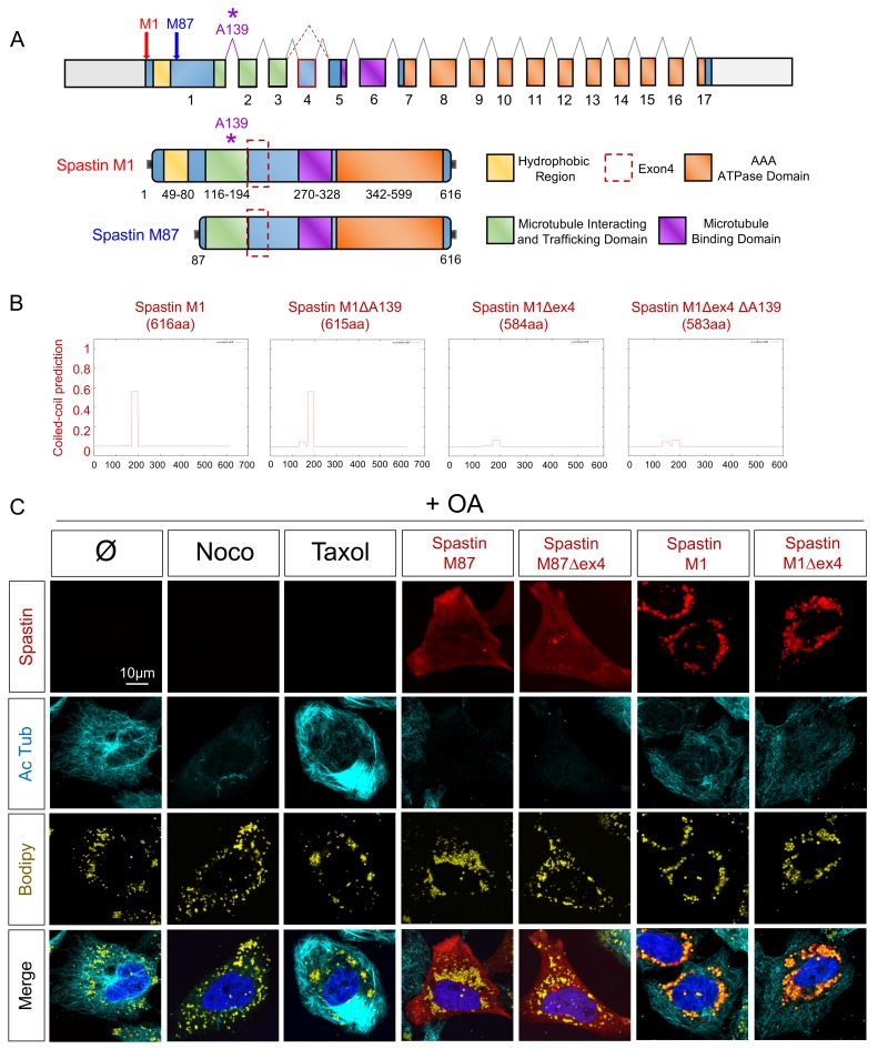
(A) Schematic representation of human Spastin splice variants. (B) Coiled-coil prediction in Spastin isoforms (designed from https://embnet.vital-it.ch/software/COILS_form.html). X-axis corresponds to the amino-acids sequence. (C) Confocal microscopy images of HeLa cells overexpressing Spastin splice variants treated with oleic acid (OA) for 18h. Nocodazole (Noco) and Taxol administration (12h) serve as control conditions. Cherry-tagged Spastin appears in red. Acetylated Tubulin labeling corresponds to microtubules (cyan), Bodipy to LDs (yellow) and Hoechst to nucleus (blue). See also S2 Fig.

Acknowledgments
This image is the copyrighted work of the attributed author or publisher, and ZFIN has permission only to display this image to its users. Additional permissions should be obtained from the applicable author or publisher of the image. Full text @ PLoS Genet.