
Image
|
Figure Caption
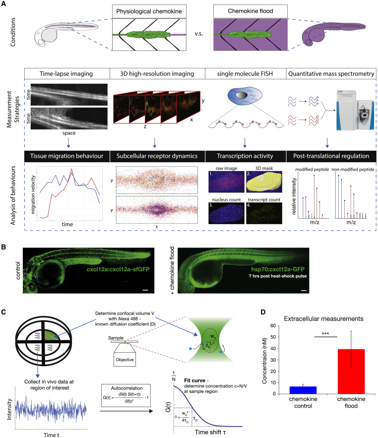
Fig. 1 An Experimental Framework to Study Robustness of Tissue Migration in Response to Changing Chemokine Concentration In Vivo (A) Experimental adaptation framework. (B) cxcl12a:cxcl12a-tagRFP-sfGFP (left) and hsp70:cxcl12a-GFP (right) embryos at 7 h after heat-shock induction. Scale bars, 100 μm. (C) Schematic for image acquisition and FCS analysis (see STAR Methods). (D) Bar charts (mean and standard deviation) of inferred in vivo chemokine concentrations in control (n = 50) and chemokine flood (n = 42) at 7 h post-induction. Statistics: ∗∗∗p < 0.001.
Acknowledgments
This image is the copyrighted work of the attributed author or publisher, and
ZFIN has permission only to display this image to its users.
Additional permissions should be obtained from the applicable author or publisher of the image.
Reprinted from Developmental Cell, 52(4), Wong, M., Newton, L.R., Hartmann, J., Hennrich, M.L., Wachsmuth, M., Ronchi, P., Guzmán-Herrera, A., Schwab, Y., Gavin, A.C., Gilmour, D., Dynamic Buffering of Extracellular Chemokine by a Dedicated Scavenger Pathway Enables Robust Adaptation during Directed Tissue Migration, 492-508.e10, Copyright (2020) with permission from Elsevier. Full text @ Dev. Cell