
|
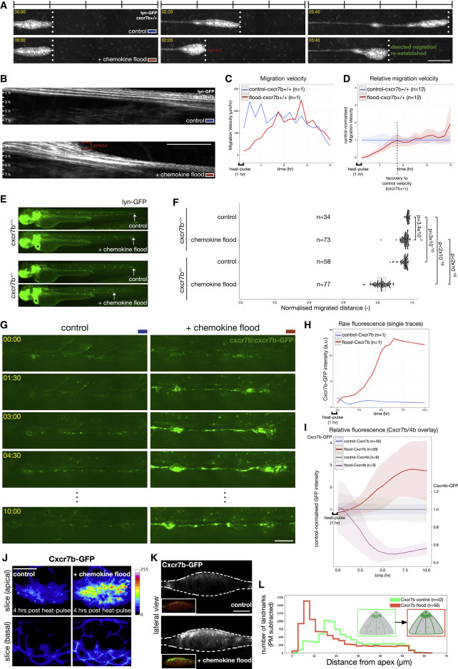
Fig. 2 Tissue Migration Rapidly Adapts to a Chemokine Flood, a Response that Is Sensitive to Cxcr7b Levels (A and B) Time lapse of lateral line migration (cldnB:lyn-GFP) in cxcr7b+/+ embryos in control (top: temperature shifted without hsp70:mCherry-cxcl12a transgene) and chemokine flood (bottom: after induction of hsp70:mCherry-cxcl12a) (A) and corresponding kymographs (B). Scale bars, 100 μm (A) and 50 μm (B). Timescale 1 h per bar. See Video S1A. (C) Lateral line migration velocity over time (h post-heat shock); control (blue) and chemokine flood (red). (D) Migration velocity in chemokine flood (n = 12, red) relative to the mean of control (n = 12, blue). Shadings, standard deviations. (E) Migration in cxcr7b+/+ or cxcr7b+/− embryos 24 h after chemokine flood. hsp70:mCherry-cxcl12a transgenics distinguished by the green heart marker (clmc2:GFP, asterisk). (F) Boxplot, median, and quantiles of normalized distance migrated. Statistics: p values indicated on the plot. (G) Time lapse of the Cxcr7b-GFP-expressing lateral line in control (left) or chemokine flood (right). Scale bar, 50 μm. See Video S2. (H) Cxcr7b-GFP fluorescence intensity over time, control (blue) and chemokine flood (red). (I) Cxcr7b-GFP and Cxcr4b-GFP intensities in chemokine flood relative to the mean of the control over time. Cxcr7b-GFP n = 30 and 29 and Cxcr4b-GFP n = 9 and 9. (J) Images of Cxcr7b-GFP in control (left) or chemokine flood (right). Single optical slices allow intensity comparison (calibration bar, signal intensity). Scale bar, 10 μm. (K) Lateral views of (J). Insets: Cxcr7b-GFP with PM label (krt15:lyn-RFP). Scale bar, 10 μm. (L) Quantification of the distance of local fluorescence point densities from tissue apex in control and chemokine flood.
Reprinted from Developmental Cell, 52(4), Wong, M., Newton, L.R., Hartmann, J., Hennrich, M.L., Wachsmuth, M., Ronchi, P., Guzmán-Herrera, A., Schwab, Y., Gavin, A.C., Gilmour, D., Dynamic Buffering of Extracellular Chemokine by a Dedicated Scavenger Pathway Enables Robust Adaptation during Directed Tissue Migration, 492-508.e10, Copyright (2020) with permission from Elsevier. Full text @ Dev. Cell