
|
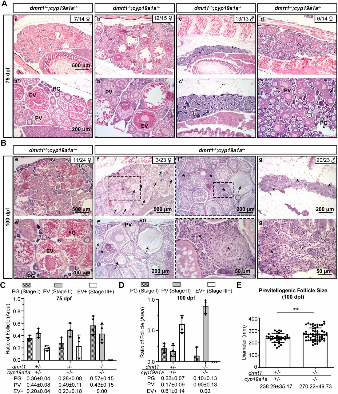
Fig. 2 Phenotype analysis of female zebrafish in different genotypes. (A,B) Histological examination of gonads in different genotypes of dmrt1;cyp19a1a at 75 dpf (A) and 100 dpf (B). PG, primary growth; PV, pre-vitellogenic; EV, early vitellogenic; FG, full grown. Arrows indicate atresic/degenerating follicles. Asterisks indicate stromal cells. (C,D) Ratios of PG, PV and vitellogenic (EV+) follicle areas in different genotypes at 75 dpf (C) and 100 dpf (D). Data are mean±s.d. The exact values are shown under the x-axis. (E) The size of PV follicles in controls and double mutants (dmrt1−/−;cyp19a1a−/−) at 100 dpf. Each dot represents the average diameter of PV follicles with visible germinal vesicles from each fish (quantified from three different sections). Data are mean±s.d. and the exact values are shown under the x-axis. Statistical significance was revealed by two-tailed unpaired Student's t-test (**P<0.01).