
Image
|
Figure Caption
Figure 1-S1
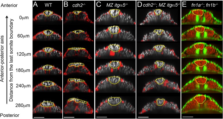
Reduction of cell-ECM interactions leads to precocious neural tube convergence and rescues cdh2 mutant neural tube convergence defects.
Each column shows a series of transverse sections in 12–14 somite stage embryos along the anterior-posterior axis of the tailbud starting from the last somite boundary (0 μm) in WT (A), cdh2-/- mutants (B), MZ itgα5-/- mutants (C), cdh2-/-; MZ itgα5-/- mutants (D), and fn1a-/; fn1b-/-mutants. (A–D) Immunostaining for FN (red) and nuclei labeling (grey). (E) F-actin labeling (green), and nuclei labeling (red). Yellow dotted lines delineate neural tube contours. Dorsal is to the top. Scale bars = 70 μm.
Acknowledgments
This image is the copyrighted work of the attributed author or publisher, and
ZFIN has permission only to display this image to its users.
Additional permissions should be obtained from the applicable author or publisher of the image.
Full text @ Elife