
Fig. 8
- ID
- ZDB-IMAGE-200323-24
- Genes
- Publication
- Caceres et al., 2019 - Frizzled 4 regulates ventral blood vessel remodeling in the zebrafish retina
- All Figures
- Figures for Caceres et al., 2019
|
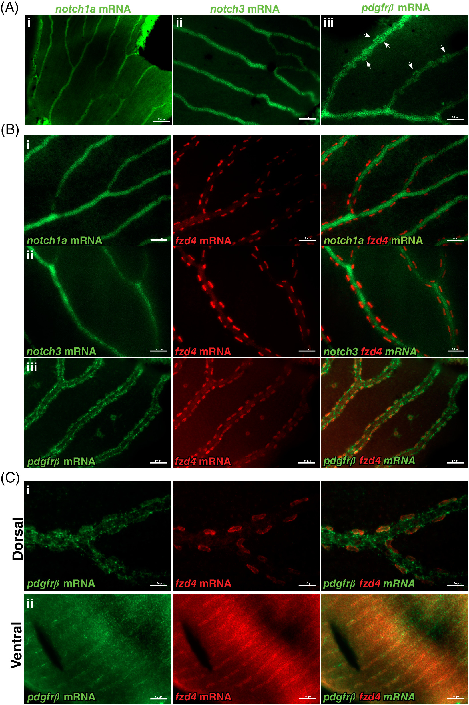
Fig. 8 fzd4 expression is detected in pericytes. Fluorescence detection of notch1a, pdgfrß, and notch3 (green) and fzd4 mRNA (red) in, A‐B, control and C, fzd4 mutant retinal vasculature. A, In control fish, strong localization of i) notch1a and ii) notch3 expression is detected throughout the retinal vasculature but not in pericytes. Similarly, in A‐iii, strong pdgfrß expression is detected throughout the retinal vasculature, with lower levels of pdgfrß expression along and within pericytes, which are localized at the outer border of the blood vessel (arrowheads). B, Double‐RNA staining of i, notch1a, ii, notch3, and iii, pdgfrß mRNA (green) with fzd4 mRNA (red), show co‐localization of fzd4 only with pdgfrß mRNA in the pericytes. C, Co‐localization of fzd4 with pdgfrß mRNA in the pericytes is only detected on the i, dorsal but not on the ii, ventral side of fzd4 mutant retinas. Scale bar in, A‐i, is 100 μm, and in, A‐ii and A‐iii, is 50 μm Scale bar in, B, i‐iii is 50 μm. Scale bar in, C‐i, is 20 μm and in, C‐ii, is 50 μm