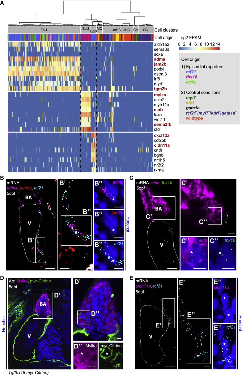
IMAGE

Figure 3

ID
ZDB-IMAGE-200318-10
Genes
Antibodies
Source
Figures for Weinberger et al., 2020
Image
Figure Caption

Figure 3

Transcriptionally Distinct Epicardial Subpopulations Epi1–3 Localize to Different Regions of the Developing Heart

(A) Marker gene expression in Epi1–3. Cells were clustered in an unsupervised manner (columns). Color key indicates range of log2 transformed FPKM values. Genes analyzed further are highlighted in red. CM, cardiomyocyte; eHC, erythroid hematopoietic cell; mHC, myeloid hematopoietic cell; NC, neural cell; and MC, mesenchymal cell.

(B) mRNA staining of the Epi1 markers adma (magenta), jam2b (red), and tcf21 (cyan) in a 5 dpf heart. (B′ and B″) A nucleus (asterisk) in the epicardial region surrounded by adma, jam2b, and tcf21.

(C) mRNA staining of the Epi2 marker elnb (magenta) and tbx18 (green) at 5 dpf. (C′ and C″) Overlap of elnb and tbx18 in the BA.

(D) Antibody staining of Mylka and tbx18:myr-Citrine at 5 dpf. (D′ and D″) Overlap of Mylka and myr-Citrine in the BA.

(E) mRNA staining of the Epi3 marker cldn11a (magenta) and tcf21 (cyan) at 5 dpf. (E′ and E″) Two nuclei (asterisks) in the epicardial region between BA and atrium in close proximity to cldn11a and tcf21. Scale bars: 20 μm in (B)–(E) and 5 μm in (B′)–(E′) and (B″)–(E″). Color channels were adjusted separately for brightness and contrast. (B)–(E) are single optical sections. V, ventricle; BA, bulbus arteriosus.

Figure Data
Acknowledgments
This image is the copyrighted work of the attributed author or publisher, and ZFIN has permission only to display this image to its users. Additional permissions should be obtained from the applicable author or publisher of the image.

Reprinted from Developmental Cell, 52(5), Weinberger, M., Simões, F.C., Patient, R., Sauka-Spengler, T., Riley, P.R., Functional Heterogeneity within the Developing Zebrafish Epicardium, 574-590.e6, Copyright (2020) with permission from Elsevier. Full text @ Dev. Cell