
Figure 2—figure supplement 6.
- ID
- ZDB-IMAGE-200304-9
- Publication
- Shahar et al., 2020 - Large-scale cell-type-specific imaging of protein synthesis in a vertebrate brain
- All Figures
- Figures for Shahar et al., 2020
|
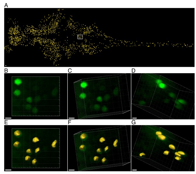
Figure 2—figure supplement 6.
Click chemistry was performed to label nascent proteins. Antibody staining for CFP was performed to detect cells expressing CFP (and MetRSL270G) across the larvae. Individual neurons were segmented in 3D according to the CFP signal, before measuring nascent proteins (in the segmented ROIs). (