
|
Figure 2
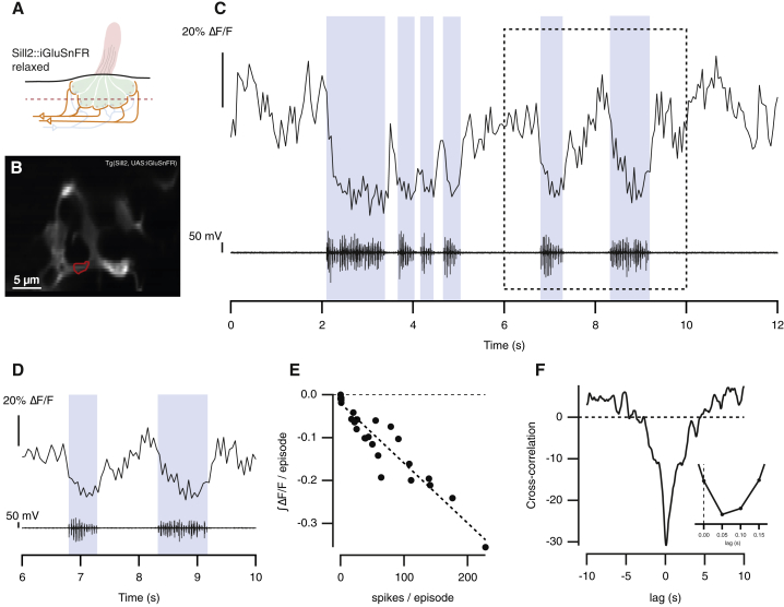
Spontaneous Release of Glutamate from Hair Cells Is Suppressed during Fictive Swimming
(A) Experiments were carried out in “relaxed” mutants that express the glutamate reporter iGluSnFR in afferent neurons (
(B) A representative hair cell synapse outlined in red.
(C) Spontaneous glutamate release from the synapse in (B) (top) and motor neuron activity measured simultaneously (bottom). Blue areas indicate bursts of fictive swimming.
(D) Magnified view of boxed area in (C). The maximum suppression of glutamate release was similar for each burst of motor activity.
(E) Relationship between the number of spikes in a burst and the negative integral of the iGluSnFR signal from the neuromast depicted in (B) (n = 28 swimming episodes; r = −0.95).
(F) Cross-correlation of iGlusnFR signal and the spike train in the motor nerve (down sampled to match imaging frequency). The inset shows that the maximum degree of anti-correlation occurred at a delay of 50 ms, indicating that the iGluSnFR signal fell within one frame interval of a spike in the motor nerve.
See also