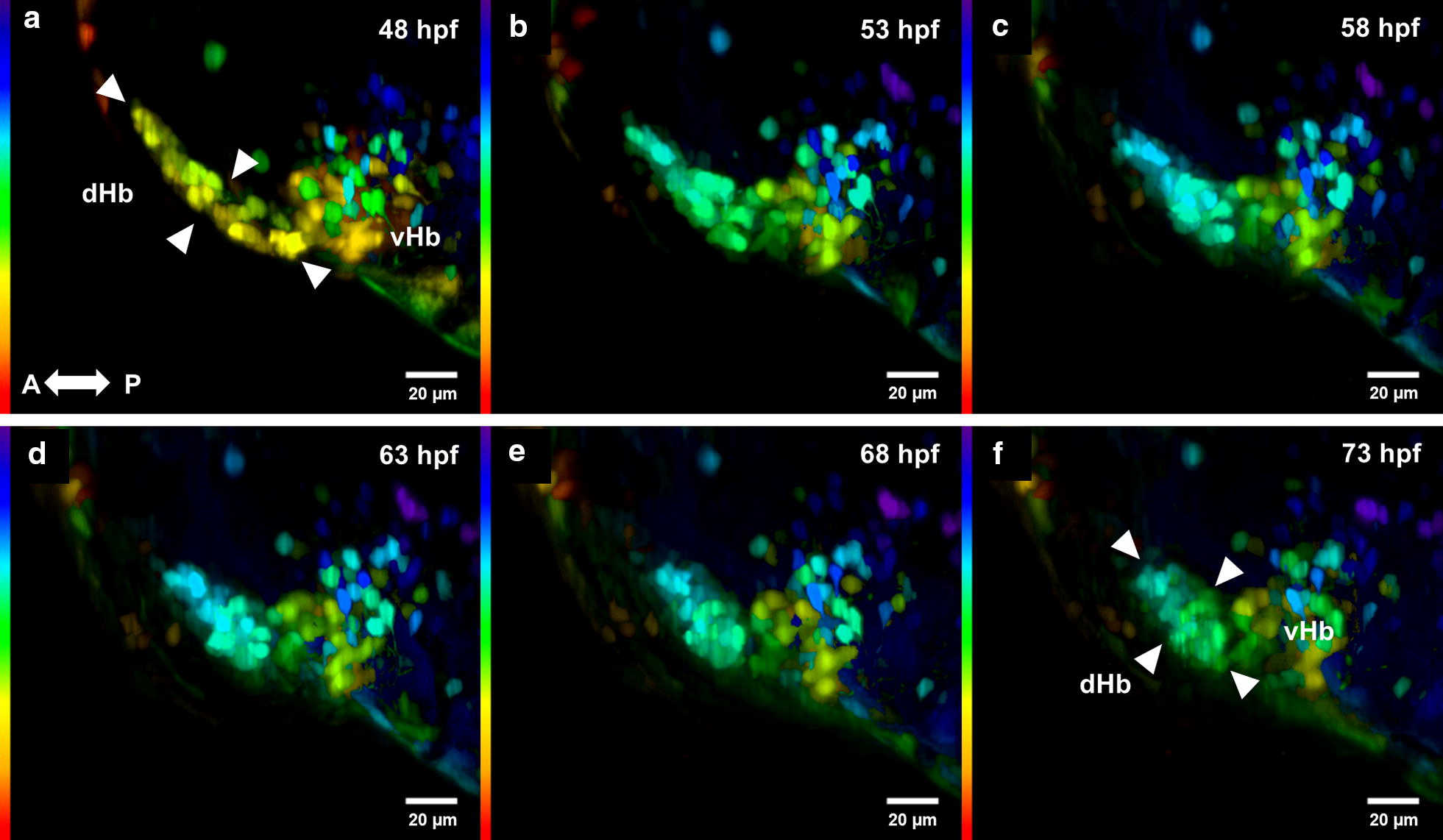
IMAGE

Fig. 3

ID
ZDB-IMAGE-200112-22
Source
Figures for Agudelo-Dueñas et al., 2020
Image
Figure Caption

Fig. 3

Development of the habenula from 48 to 73 hpf. Development of the Hb complex followed through time-lapse imaging. Cells within the dorsal habenula (dHb) aggregate progressively during development from an elongated shape to form a nucleus as indicated by the white arrowheads. The dHb is observed to be located more dorsally with respect to the vHb. Images from af are MIP color coded for a depth of 250 µm, purple and blue being more dorsal, and red and orange more ventral. Original stacks were cropped and aligned using the FiJi plugin Linear Stack Alignment with SIFT [20]. Gamma was adjusted to a value of 0.75. All time points are dorsal views. A, anterior, P, posterior

Acknowledgments
This image is the copyrighted work of the attributed author or publisher, and ZFIN has permission only to display this image to its users. Additional permissions should be obtained from the applicable author or publisher of the image. Full text @ BMC Res. Notes