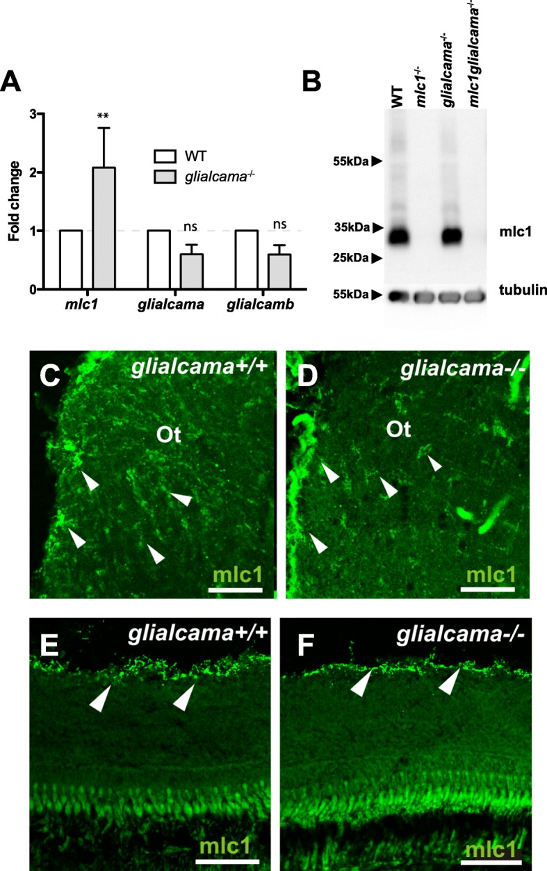
IMAGE

Fig. 4

ID
ZDB-IMAGE-191230-692
Genes
Antibodies
Source
Figures for Pérez-Rius et al., 2019
Image
Figure Caption

Fig. 4

mlc1 expression and localization in glialcama−/− zebrafish. a Quantitative real-time PCR to determine levels of glialcama, glialcamb and mlc1 messenger RNA in the brain of glialcama−/− zebrafish. Bars, relative expression levels compared with WT sibling; error bars, s.e. (n ≥ 3). **P < 0.01 (vs. wild-type, two-way ANOVA) b Comparison of mlc1 protein levels in brain of wild-type (WT), mlc1−/−, glialcama−/− and mlc1glialcama−/− zebrafish by Western blots of extracts from 5-months-old zebrafish. Western blot is representative of three independent experiments. Tubulin served as a loading control. c, d mlc1 expression (arrowheads) observed in the optic tract of both wild type (c) and glialcama−/− (d) brains. e, f mlc1 expression is restricted to the inner limiting membrane of the retina (arrowheads) both in wild type (e) and glialcama−/− (f). Scale bars: 50 μm (c, d); 60 μm (e, f)

Figure Data
Acknowledgments
This image is the copyrighted work of the attributed author or publisher, and ZFIN has permission only to display this image to its users. Additional permissions should be obtained from the applicable author or publisher of the image. Full text @ Orphanet J Rare Dis