IMAGE

FIG 3

ID
ZDB-IMAGE-191230-203
Source
Figures for Burggraaf et al., 2019
Image
Figure Caption

FIG 3

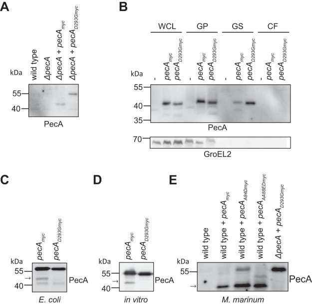
PecA is subjected to self-cleavage. (A) Expression of PecAmyc and PecAD293Gmyc in M. marinumΔpecA was detected by immunoblotting of whole-cell lysate. (B) Subcellular localization and secretion analysis of M. marinum wild type (indicated with “-”) or wild type expressing PecAmyc or PecAD293Gmyc. WCL, whole-cell lysate; GP, Genapol-treated pellet (membrane associated and intracellular); GS, Genapol supernatant (membrane associated); CF, culture filtrate. GroEL2 was probed as a cytosolic control. (C) Immunoblot analysis of PecAmyc and PecAD293Gmyc expressed in E. coli (whole-cell lysate). (D) Immunoblot analysis of in vitro translation of PecAmyc and PecAD293Gmyc. (E) Immunoblot analysis of M. marinum wild type; wild type expressing PecAmyc, PecAA84Dmyc, or PecAAA88EDmyc; and ΔpecA mutant expressing PecAD293Gmyc (whole-cell lysate). The arrow indicates the fully cleaved form of PecA. Proteins were detected by their C-terminal Myc tag with an anti-Myc antibody.

Acknowledgments
This image is the copyrighted work of the attributed author or publisher, and ZFIN has permission only to display this image to its users. Additional permissions should be obtained from the applicable author or publisher of the image. Full text @ MBio