Figure Caption
Fig 4
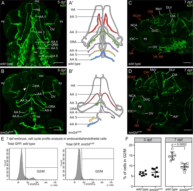
<italic>kmt2d</italic><sup><italic>zy59</italic></sup> mutants fail to develop AAs and exhibit misspatterned cephalic vasculature.(A-B) Ventral view of vasculature in wild-type tg(kdrl:GFP) sibling (A) and kmt2dzy59;tg(kdrl:GFP) mutants at 5 dpf. Cephalic vascular architecture in kmt2dzy59 embryos was abnormal with reduced elongation in the anteroposterior body axis, loss of bilateral symmetry, and reduced HA (A, B). In kmt2dzy59 mutants, AA1 (A, B) was shorter with minimum elongation towards the anterior area of the embryo, the AA3 (A, B) was rudimentary,whereas AA4−6 were reduced or absent (A, B). Mouth and eyes in kmt2dzy59 showed primitive characteristics with differences in the thickness of the OV in particular (A–B). Note the abnormal endocardial component of the heart (A, B; he). Aʹ and B are simplified cartoons of the main differences in the AAs development in wild-type (Aʹ) and mutant (Bʹ) backgrounds. Branchial vasculature loops were removed to allow better visualization of aortic arch points of origin. (C-D) Lateral view of cephalic vasculature in wild-type tg(kdrl:GFP) sibling and kmt2dzy59;tg(kdrl:GFP) mutants at 7 dpf. kmt2dzy59 exhibited a complete absence of the vascular loops associated with AAs 3–6 with only a rudiment of the AA3 present (D, AA*). The general cranial vascular network was mispatterned with a particular strong impact in the CtA, ACeV, and VA (D; in orange). In kmt2dzy59 mutant, most vessels had reduced lumens with the exception of the NCA (C, D), DVC (C, F) and IOC (C, F) that show thicker vessel diameter. Note the reduced HA from a lateral view (D, HA in orange; scale bars = 100 μm). (E) Cell cycle profile analysis by FACS for 7 individual embryos per genotype at 7 dpf. The gates set up for nuclear staining (DAPI) in kdrl positive cells (endothelial and endocardial cells) are shown. (F) Cell cycle profiles for Kdrl positive cells in kmt2dzy59 mutants showed no significant difference in the percentage of G2/M cells at 3 dpf. In contrast, at 7 dpf, there was a significant decrease in number of dividing cells in kmt2dzy59 mutants when compared with wild-type siblings. Unpaired two-tailed t test, p = 0.407 n.s. for 3 dpf; p = 0.0002 for 7 dpf, n = 7 per genotype. Values for each data point can be found in S1 Data. AA, aortic arch; AA1, mandibular arch; AA2, hyoid arch; AA3, first branchial arch; AA4, second branchial arch; AA5 third branchial arch; AA6, fourth branchial arch; ACeV, anterior cerebral vein; CtA, central artery; DCV, dorsal ciliary vein; DLV, dorsal longitudinal vein; dpf, days post fertilization; ey, eye; FACS, fluorescent activated cell sorting; GFP, green fluorescent protein; HA, hypobranchial artery; kdrl, kinase insert domain receptor like; IOC, inner optic circle; MsV, mesencephalic vein; NCA, nasal ciliary artery; ORA, opercular artery; OV, optic vein; VA, ventral aorta.
Acknowledgments
This image is the copyrighted work of the attributed author or publisher, and
ZFIN has permission only to display this image to its users.
Additional permissions should be obtained from the applicable author or publisher of the image.
Full text @ PLoS Biol.