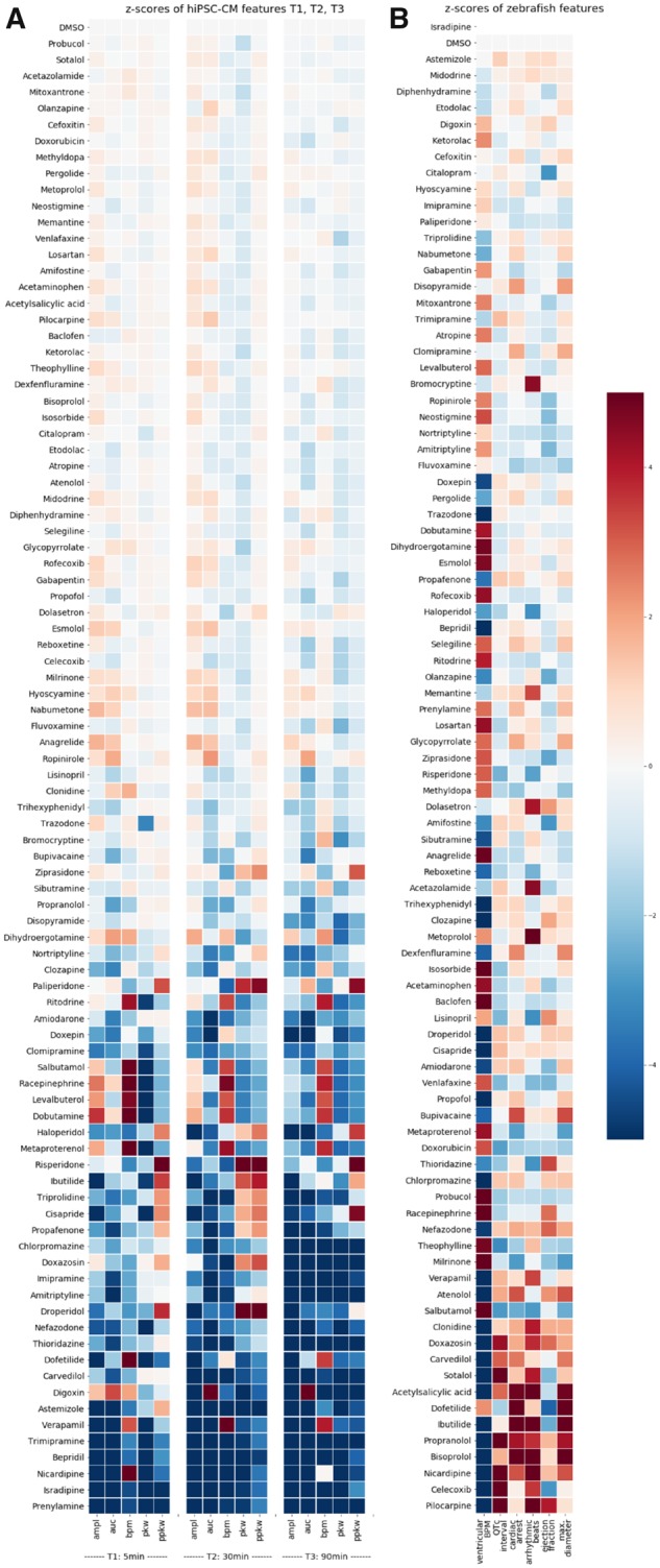
IMAGE

Figure 2.

ID
ZDB-IMAGE-191230-1035
Source
Figures for Dyballa et al., 2019
Image
Figure Caption

Figure 2.

Heatmap of the z-scores of human induced pluripotent stem cell cardiomyocyte (hiPSC-CM) and zebrafish features. A, The z-scores of features measured by fluorometric imaging plate reader (FLIPR) in hiPSC-CMs at 5 min (T1), 30 min (T2), and 90 min (T3) after compound addition are shown for each compound of the library: amplitude (ampl), area under curve (auc), beats per minute (bpm), peak width (pkw), and peak width at 10% amplitude (ppkw). The z-scores were computed from the effects determined by the statistical model for each compound and each time point. B, The z-scores of numerical features in zebrafish larvae 4 h after compound addition quantified by ZeCardio and analyzed by the statistical model are shown for each compound of the library. Six features are shown: ventricular beats per minute (ventricular BPM), QTc interval (QTc), longest beat (cardiac arrest), percentage of arrhythmic beats (arrhythmic beats), estimated ejection fraction (ejection fraction), and the maximum ventricular diameter (max. diameter). All cells are color-coded according to their z-score (color bar on the right hand side) and compounds in (A) or (B) are ordered according to the sum of absolute z-scores for each compound from bottom to top.

Acknowledgments
This image is the copyrighted work of the attributed author or publisher, and ZFIN has permission only to display this image to its users. Additional permissions should be obtained from the applicable author or publisher of the image. Full text @ Toxicol. Sci.