
Image
|
Figure Caption
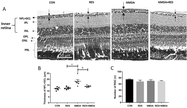
Fig. 5
Resveratrol prevents from NMDA-induced thicker NFL
A: H&E staining of paraffin sections (4 µm) from representative retinas treated by I.M. in distilled water (control), 50 mg/L resveratrol, 100 µmol/L NMDA, or 50 mg/L resveratrol+100 µmol/L NMDA. The dark arrows point out the thickness of each layer. B: The thickness of the NFL+GCL for each treatment group; C: The retinal ganglion cell number for each treatment group. Error bars represent standard error of the mean (±SEM); n=6 (unpaired t-test, bP<0.01 and cP<0.001). Original magnification is 40×. Scale bar, 50 µm.
Acknowledgments
This image is the copyrighted work of the attributed author or publisher, and
ZFIN has permission only to display this image to its users.
Additional permissions should be obtained from the applicable author or publisher of the image.
Full text @ Int J Ophthalmol