
|
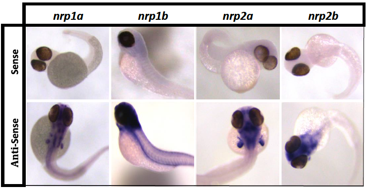
Fig. S2
nrp riboprobes validation.
In situ hybridization of TraNac transgenic zebrafish embryos 48 hours post fertilization (hpf) with nrp sense riboprobes (upper row) and nrp anti-sense riboprobes (lower row). Anti-sense riboprobes differential staining patterns were compared to previous reports (43) to confirm specific nrp isoform detection. All neuropilin isoforms are observed in the brain with additional differential expression patterns observed between different isoforms. Nrp1a is observed in the fin buds and otic vesicles, nrp1b is expressed in the dorsal aorta and intersegmental vessels, nrp2a is observed in the hind brain and fin buds, whereas nrp2b is largely restricted to the brain and hind brain, n ≥ 8.