
IMAGE
Fig. S2
- ID
- ZDB-IMAGE-191007-17
- Publication
- Nicholas et al., 2019 - Temporal characterization of optic fissure basement membrane composition suggests nidogen may be an initial target of remodeling
- All Figures
- Figures for Nicholas et al., 2019
Image
|
Figure Caption
Fig. S2
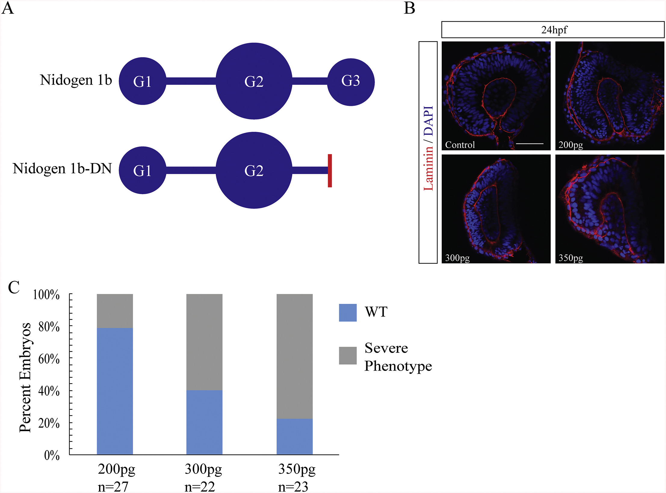
Nidogen dominant negative mRNA injections result in ocular malformation. A) Schematic diagram of nidogen functional domains and the strategy for generating a dominant negative nidogen construct by truncating nidogen 1b of the G3 domain. Red bar indicates deletion point of globular domain 3 in nid1b-DN. B) Retinal phenotypes in 24hpf nid1b-DN mRNA injected embryos characterized using laminin IHC (red), and DAPI (blue). Single confocal sections are depicted. Scale bar = 50 μm. C) Increasing concentration of nid1b-DN mRNA resulted in higher percentage of ocular morphological defects observed at 24hpf.
Acknowledgments
This image is the copyrighted work of the attributed author or publisher, and
ZFIN has permission only to display this image to its users.
Additional permissions should be obtained from the applicable author or publisher of the image.
Reprinted from Developmental Biology, 452(1), Nicholas, C., Weaver, M., Piedade, W.P., Vocking, O., Famulski, J.K., Temporal characterization of optic fissure basement membrane composition suggests nidogen may be an initial target of remodeling, 43-54, Copyright (2019) with permission from Elsevier. Full text @ Dev. Biol.