
Fig. 5
- ID
- ZDB-IMAGE-191007-11
- Antibodies
- Publication
- Nicholas et al., 2019 - Temporal characterization of optic fissure basement membrane composition suggests nidogen may be an initial target of remodeling
- All Figures
- Figures for Nicholas et al., 2019
|
Fig. 5
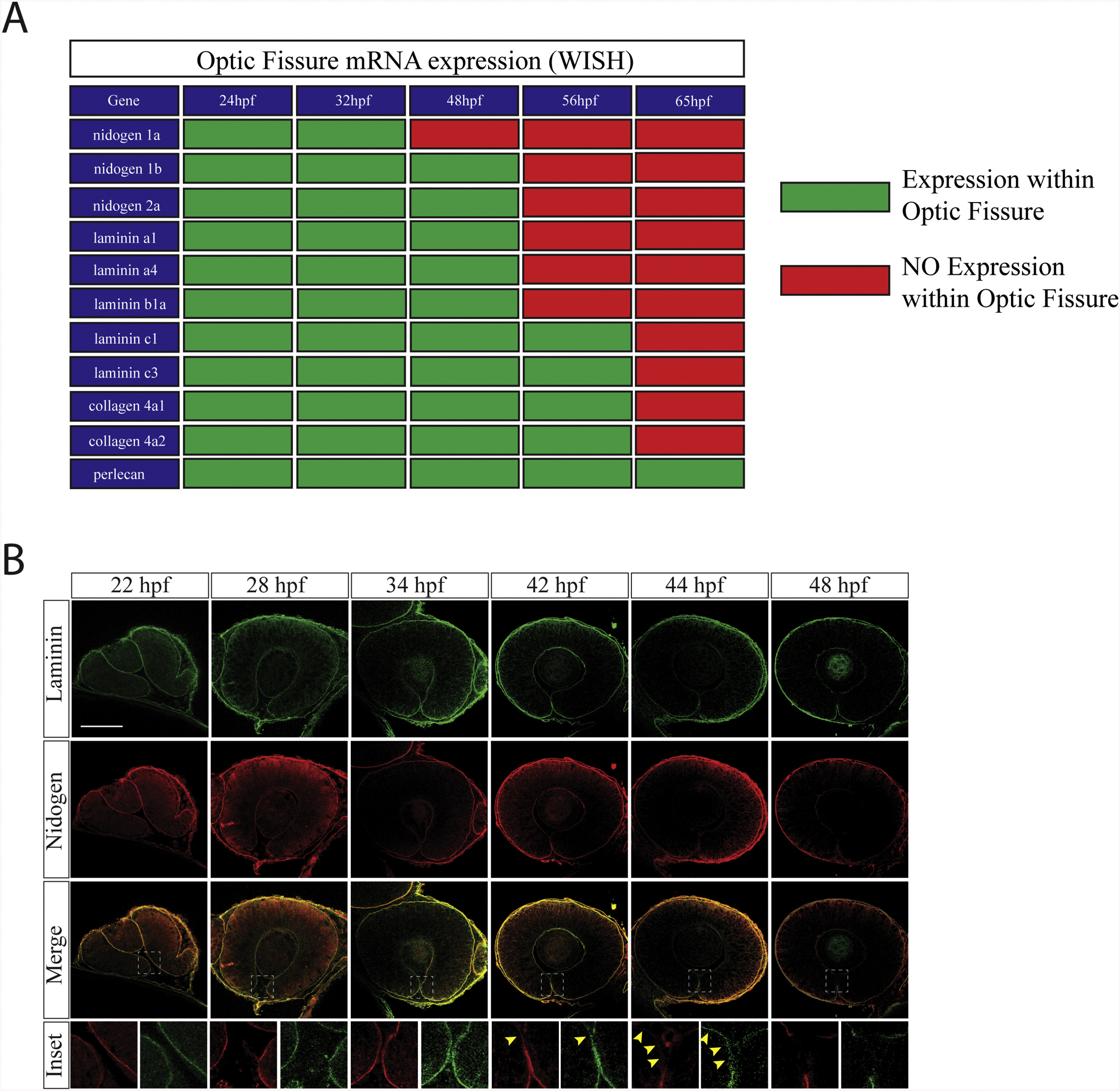
Nidogen is down-regulated prior to laminin during optic fissure fusion. A) Summary of core basement membrane component expression within the optic fissure as observed by WISH (Fig. 1, Fig. 2, Fig. 3, Fig. 4) between 24 and 65hpf. Green indicates expression (WISH signal), red indicates absence of expression (lack of WISH signal). B)Immunohistochemistry analysis for remodeling of laminin (green) and nidogen 1 (red) within the optic fissure during retinal morphogenesis(22-48hpf). At early time points, ∼22-40hpf, laminin and nidogen signal is found to co-localize in the optic fissure BM. Starting at 42hpf nidogen signal in the optic fissure is reduced prior to that of laminin (yellow arrow heads). By 48hpf both laminin and nidogen are absent in the regions of the fissure where fusion has completed. Scale bar = 50 μm.
Reprinted from Developmental Biology, 452(1), Nicholas, C., Weaver, M., Piedade, W.P., Vocking, O., Famulski, J.K., Temporal characterization of optic fissure basement membrane composition suggests nidogen may be an initial target of remodeling, 43-54, Copyright (2019) with permission from Elsevier. Full text @ Dev. Biol.