
|
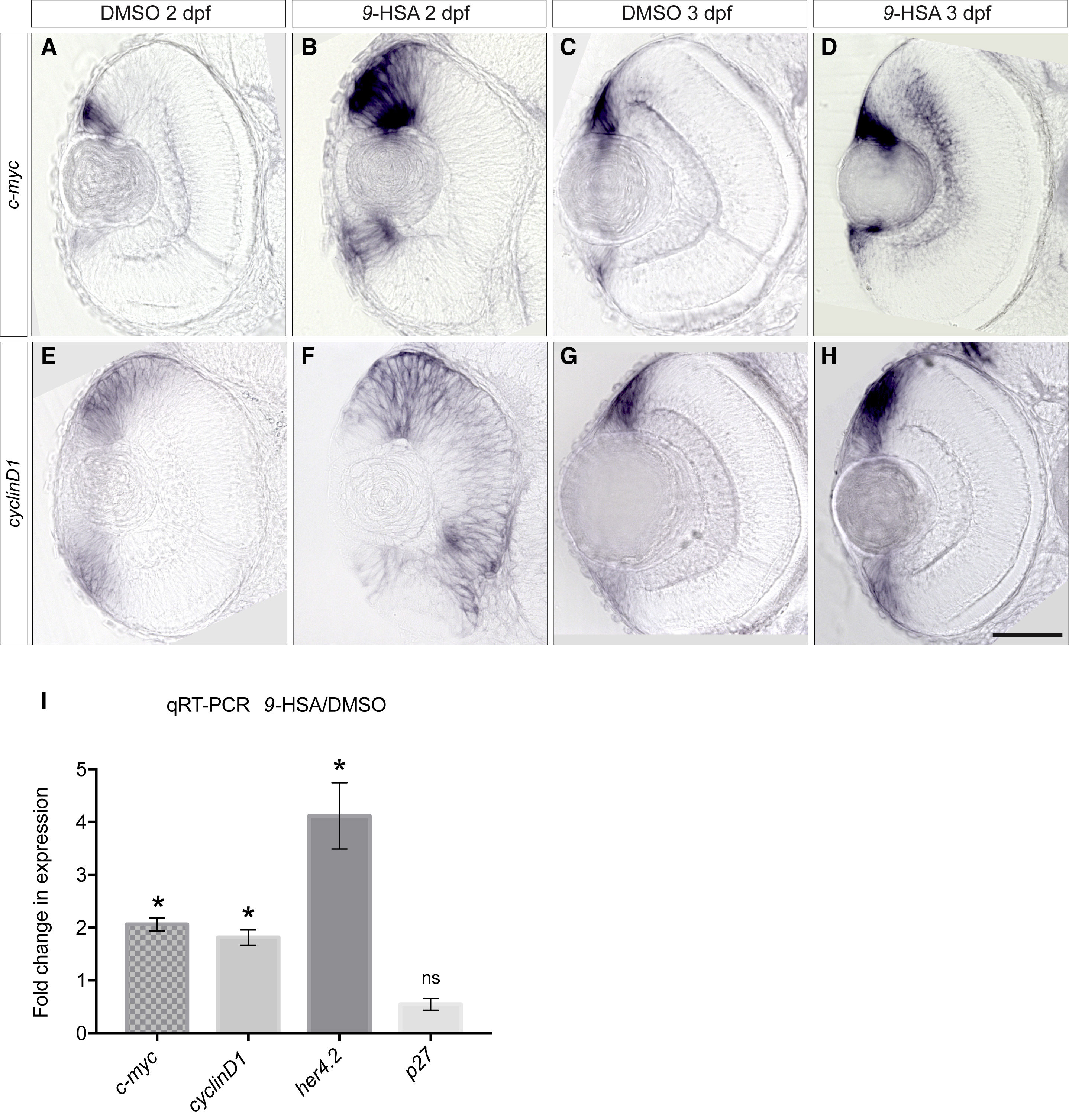
Fig. 4
9-HSA Induces RPCs Proliferation via the Activation of Canonical Wnt Signaling
Expression analysis of the c-myc and cyclinD1 Wnt targets at 2 and 3 days postfertilization (dpf) in DMSO-treated (respectively, (A), (E), (C), and (G)) and 9-HSA-treated retinae (respectively (B), (F), (D), and (H)).
(A–D) Comparative in situ hybridization revealed an increased expression of c-myc in the CMZ and in the central part of the 9-HSA-treated retinae in comparison to DMSO-treated control retinae at 2 and 3 dpf.
(E–H) Similarly to c-myc, comparative analysis of cyclinD1 expression in DMSO-treated and 9-HSA-treated retinae showed an increased expression of cyclinD1 in the retinae embryos injected with 9-HSA in comparison to DMSO control retinae.
(I) Quantitative real-time PCR of c-myc, cyclinD1, her4.2 and p27expression levels in 2 dpf 9-HSA-injected embryo heads versus DMSO-injected embryo heads (n = 3 for all conditions; her4.2 fold increase of 4.117 SEM ± 0.6289, p value = 0.0384; c-myc fold increase of 2.059 SEM ± 0.1231, p value = 0.0132; cyclinD1 fold increase of 1.814 SEM ± 0.1441, p value = 0.0299; p27 fold increase of 0.5461 SEM ± 0.1119, p value = 0.0557). Statistically significant was using a Student’s t test and depicted as: ∗p value < 0.05; ns = nonsignificant. Scale bar, 50 μm.
Reprinted from Developmental Cell, 50(1), Albadri, S., Naso, F., Thauvin, M., Gauron, C., Parolin, C., Duroure, K., Vougny, J., Fiori, J., Boga, C., Vriz, S., Calonghi, N., Del Bene, F., Redox Signaling via Lipid Peroxidation Regulates Retinal Progenitor Cell Differentiation, 73-89.e6, Copyright (2019) with permission from Elsevier. Full text @ Dev. Cell