
Image
|
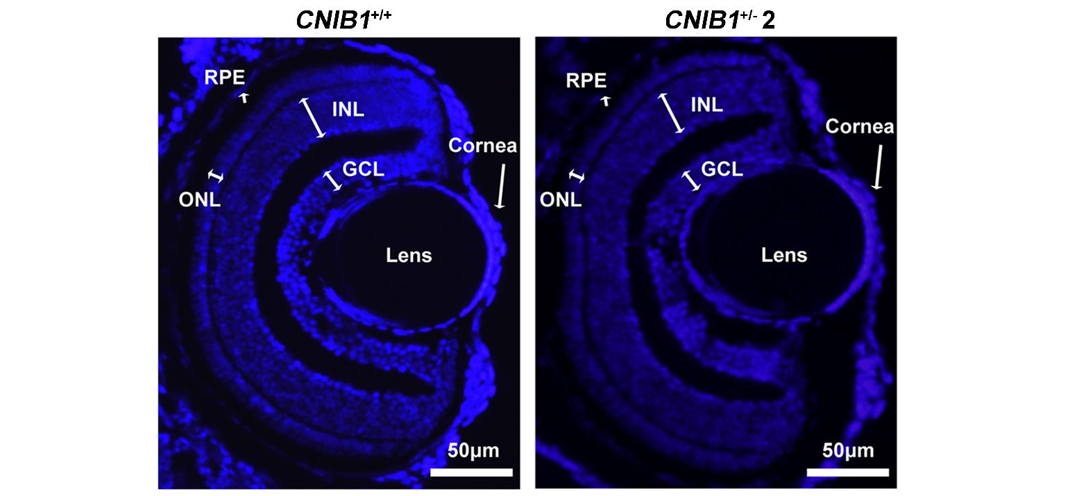
Figure Caption
Fig. S8
Representative DAPI staining images of the retinal structures shown in the zebrafish offspring at 5 dpf from CNIB1+/+ and CNIB1+/- 2 incross (n=6). Scale bars: 50 μm. GCL: ganglion cell layer; INL: inner nuclear layer; ONL: outer nuclear layer; RPE: retinal pigment epithelium.
Acknowledgments
This image is the copyrighted work of the attributed author or publisher, and
ZFIN has permission only to display this image to its users.
Additional permissions should be obtained from the applicable author or publisher of the image.
Full text @ Nucleic Acids Res.