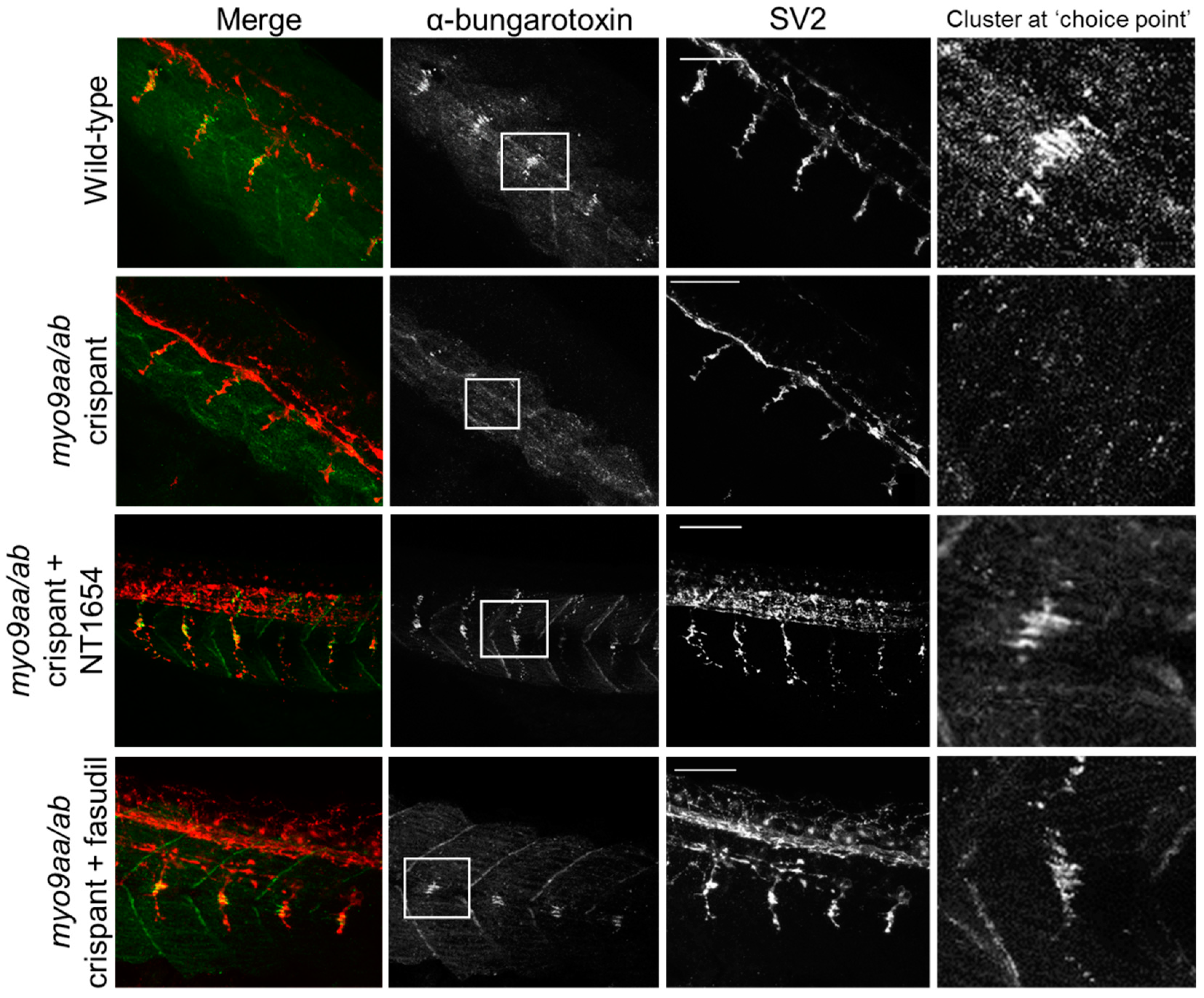
Image


                        Figure Caption


Fig. 3
Neuromuscular junction (NMJ) morphology of myo9aa/abcrispant zebrafish at 24 hpf. Representative images of NMJs in wildtype, myo9aa/ab crispant, and NT1654 (0.15 ng) or fasudil-treated (10 µm) crispant zebrafish at 24 hpf. Acetylcholine receptors stained with αBTx (green), and motor neurons detected with an antibody against SV2 (red). White boxes demark areas enlarged in the right-hand panel, showing presence of αBTx-positive choice point clusters. Scale bar = 50 µm.
                
                    
                        Figure Data


                        Acknowledgments


This image is the copyrighted work of the attributed author or publisher, and
ZFIN has permission only to display this image to its users.
Additional permissions should be obtained from the applicable author or publisher of the image.
      Full text @ Cells


        