
|
Fig. S5
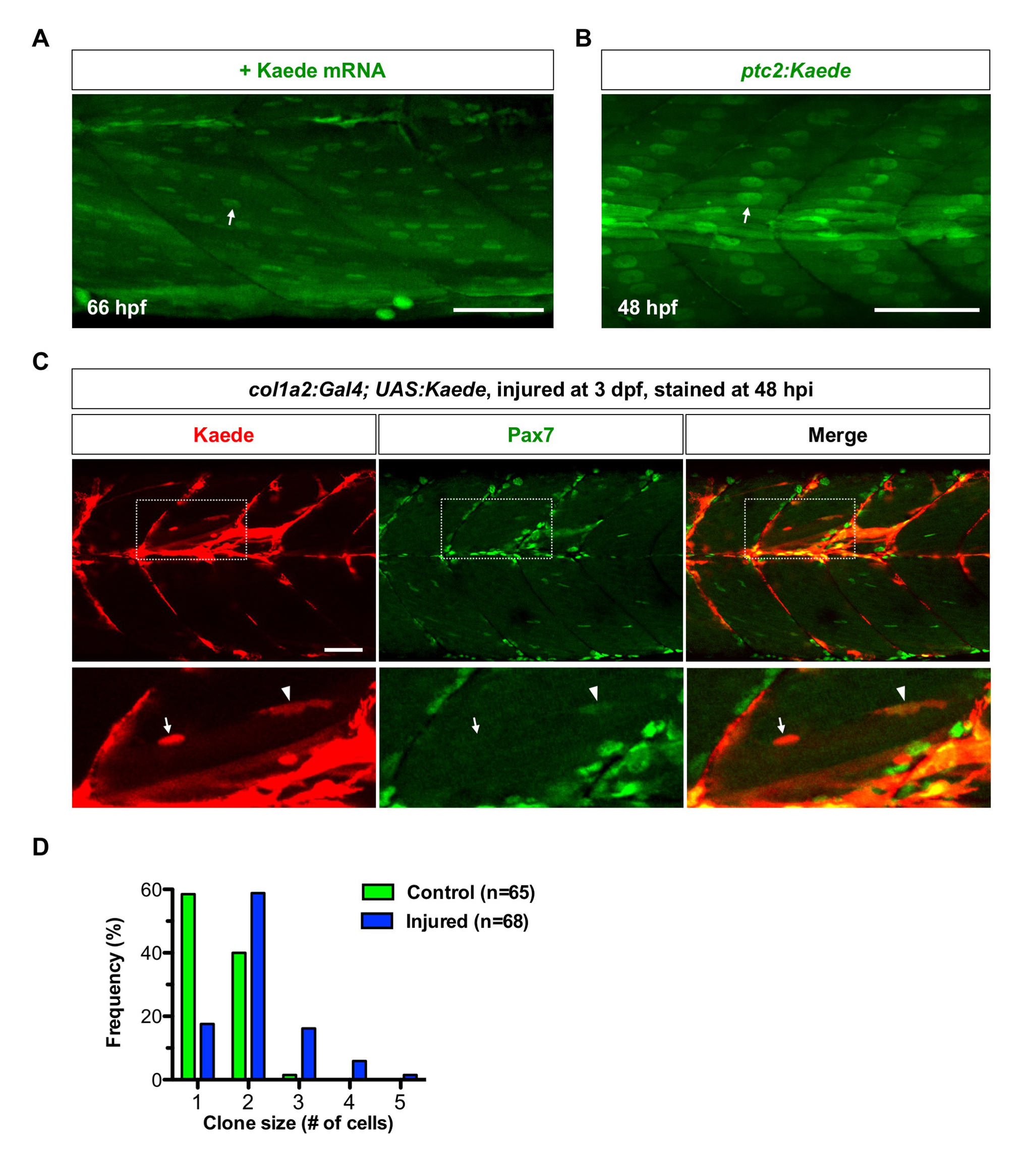
Kaede protein is preferentially localized in nuclei of muscle fibers. (A) Wildtype embryos were injected with Kaede mRNA, and imaged at 66 hpf. Kaede protein (green) is preferentially localized in the nuclei of muscle fibers (arrow). n = 40 embryos. (B) Kaede protein (green) is concentrated in the nuclei of slow myofibers (arrow) in ptc2:Kaede embryos at 48 hpf. n = 40 embryos. (C) col1a2Kaede embryos were injured at 3 dpf, and stained at 48 hpi Supplementary information with the anti-Pax7 antibody (green). Kaede+ cells (red) contributed to muscle regeneration (boxed regions). The expanded views show that newly formed muscle fiber displayed strong Kaede expression in the nucleus (arrows), which was not labelled by Pax7. By contrast, a small elongated Kaede+ cell between muscle fibers was Pax7 positive (arrowheads). n = 11 embryos. (D) Quantification of clone size in single cell clonal analysis described in Fig 5. col1a2+ MPCs under the injury condition (blue, n=68) tend to generate larger clones compared to MPCs in the wild-type condition (green, n=65). Scale bars: 50 μm.