
|
Fig. S8
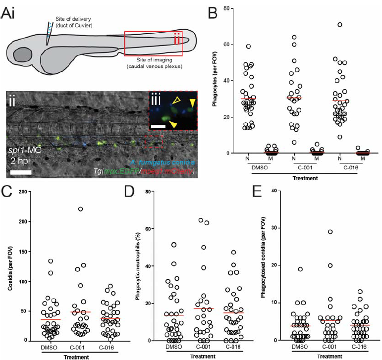
Bifunctional compounds do not enhance phagocytosis of conidia by
wild-type zebrafish neutrophils.
A) Diagram of a 72 hpf zebrafish larva indicating the site of inoculation and the site of
analysis (i). (ii) Collapsed z-stack (maximum intensity) representative image showing
conidia (blue, Hoechst) and neutrophils (green, GFP) in the caudal venous plexus of
Tg(mpx:GFP/mpeg1:mCherry) embryos injected with spi1-MO at the one-cell stage
and infected with A. fumigatus conidia at 72 hpi. Scale: 100 μm. iii) highermagnification
of neutrophils and conidia indicating extracellular conidia (open yellow
arrowhead) and examples of phagocytosis by neutrophils (filled yellow arrowheads).
Scale: 20 μm.
B) Graph shows neutrophil (N) and macrophage (M) counts in each caudal venous plexus
field of view (FOV) for spi1-MO morphant larvae injected with treated and control
conidia.
C) Graph shows conidia counts per FOV for each treatment group.
D) Graph shows the percent of neutrophils containing conidia at 2 hpi for each treatment
group.
E) Graph shows the number of phagocytosed conidia per FOV at 2 hpi for each treatment
group. Each point represents an infected larva.