
|
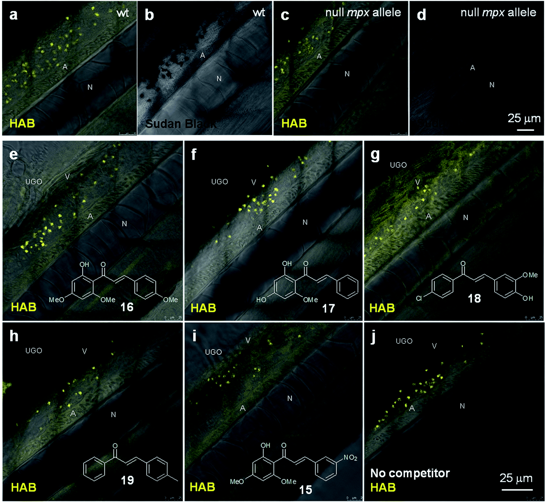
Fig. 8
HAB does not target zebrafish neutrophil myeloperoxidase, and its binding to neutrophil granules is not a general feature of chalcones. (a and c) Merged confocal fluorescence and bright-field imaging of HAB (10 μM) in live wild-type (a) or “Spotless” (NL 144_01 mutant: null mpx allele) (c) 72 hpf zebrafish larvae following excitation at 488 nm and detection in the 550–650 nm range under diffusion conditions. (b and d) SB staining of myeloperoxidase-containing neutrophil granules in bright-field imaging. (e–j) Merged confocal fluorescence and bright-field imaging of HAB (10 μM) in live wild-type 72 hpf zebrafish larvae co-treated with chalcones 15, 16 (flavokawain A), 17(cardamonin), 18 or 19 (100 μM) following excitation at 488 nm and detection in the 550–650 nm range under diffusion conditions. The yellow-orange color is indicative of the fluorescence seen with the naked eye. Single 2 μm optical sections are shown. Abbreviations used: A (aorta); N (notochord); UGO (urogenital opening); V (vein).