IMAGE

Fig 2

ID
ZDB-IMAGE-190723-574
Source
Figures for Phan et al., 2019
Image
Figure Caption

Fig 2 Reporter expression in the developing caudal fin of <italic>Tg(2P-EIand1</italic>:<italic>eGFP)</italic>.

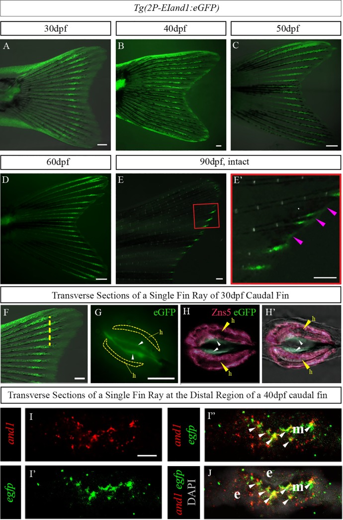
(A) Reporter expression of Tg(2P-EIand1:eGFP) (25 fish were analysed) occurs within the ectodermal tissue and fin rays along the proximal-distal axis of the developing caudal fin. It is notably brighter as it approaches the distal region at 30dpf. (B) Reporter expression occurs along the proximal-distal axis in the fin rays and is beginning to approach the distal edge of the fin where ectodermal-specific expression is present at 40dpf. (C, D) At 50dpf and 60dpf, eGFP expression in the fin rays distalizes and is present in the epithelial tissue located at the very distal regions of the fin. (E, E’) Reporter expression only occurs at the distal tips of the fin rays and at the distal edge of the interrays (pink arrowheads). (E’) close-up on panel E. (F) Transverse sections from a 30dpf caudal fin were obtained in the region indicated by the yellow dotted line. (G) Immunostaining showing eGFP expression (white arrowheads) localized deep to the hemirays (h; delineated by yellow line) (n = 6). Immunostaining for Zns5, which is a pan-osteoblast marker, labelling osteoblasts surrounding the hemirays (h; yellow arrowheads) and eGFP (small white arrowheads) in Tg(2P-EIand1:eGFP) on transverse cryosections of 30dpf larvae (n = 6). (H) eGFP is localized in layers of cells deep and adjacent to osteoblast layers lining the hemirays (h). (H’) Merge on bright field. Double fluorescence in situ hybridization (n = 12) for and1 (I) and eGFP of Tg(2P-EIand1:eGFP) (I’). (I”) Merge between red and green showing colocalization between and1 and egfp (white arrowheads). (J) Merge with DAPI showing colocalization occurring within the fin ray mesenchymal tissues. Note: Any expression observed outside of the fin ray mesenchymal compartment is background staining. n = # of fish from which sections were obtained. Scale bars: A-E, E’ = 200μm, F = 100μm, G, H-H’ = 50μm and I-I”, J = 10μm.

Acknowledgments
This image is the copyrighted work of the attributed author or publisher, and ZFIN has permission only to display this image to its users. Additional permissions should be obtained from the applicable author or publisher of the image. Full text @ PLoS One