IMAGE

Fig 1

ID
ZDB-IMAGE-190723-573
Source
Figures for Phan et al., 2019
Image
Figure Caption

Fig 1 Differential regulation of <italic>and1</italic> during larval development and adulthood.

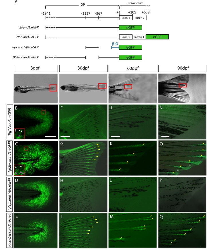
(A) Schematic representation of transgenic reporter constructs. GFP expression of four and1 reporter lines at 3dpf (B-E), 30dpf (F-I), 60dpf (J-M) and 90dpf (N-Q) showing fluorescent images merged onto bright field images. (B, B’, C, C’) confocal images (note: autofluorescence in the trunk tissue in C). Note: obtaining exactly the same plane of focus in the two transgenic lines is difficult; however, these images are simply to represent both transgenic lines have eGFP-positive ectodermal and mesenchymal cells. (B’, C’) 2x higher magnification on region boxed in red in panels B and C, respectively, showing the more elongated mesenchymal cells (pink arrowheads) in contrast to the hexagonally-shaped ectodermal cells (white arrowheads). (B, F, J, N) Tg(2Pand1:eGFP) (n = 5) expression in the ectoderm and fin ray mesenchyme fades after 30dpf. (C, G, K, O) Tg(2P-EIand1:eGFP) (n = 2) expression in the ectoderm and fin ray mesenchyme persists after 30dpf. (D, H, L, P) Tg(epi.and1-βG:eGFP) (n = 2) expression in the ectoderm is absent at 30dpf and onward; and (E, I, M, Q) Tg(2PΔepi.and1:eGFP) (n = 2) expression in the fin ray mesenchyme persists. Yellow arrowheads indicate fin ray-specific reporter expression. n = # of lines, ~25 fish/line. Scale bars = 200μm.

Acknowledgments
This image is the copyrighted work of the attributed author or publisher, and ZFIN has permission only to display this image to its users. Additional permissions should be obtained from the applicable author or publisher of the image. Full text @ PLoS One