
|
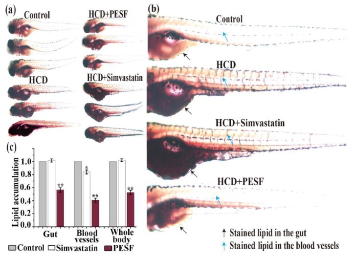
Figure 5
Comparison of PESF and simvastatin on inhibition of lipid accumulation in the zebrafish larvae model. (a) Representative photographs of zebrafish larval appearance with stainable lipids treated by PESF and simvastatin in the zebrafish model. Zebrafishes at 6 d.p.f were fed with HCD from 0 h to 24 h uninterruptedly and exposed to PESF (200 µg/mL) and simvastatin (200 µg/mL) from 12 h to 24 h and then stained by ORO at 24 h for image acquisition. For the zebrafishes in the normal group, no HCD was fed during the culture time. Conversely, the HCD group was fed with HCD for 24 h uninterruptedly. (b) Representative photographs of zebrafish larval appearance with stainable red lipids treated by PESF and simvastatin compared with the HCD group and the normal group. Black arrows point to lipids in blood vessels and blue arrows point to lipids in the gut. (c) Normalized quantification data of stainable lipid content in the zebrafish (gut, blood vessels, whole body) treated by PESF and simvastatin, correlated to microphotographs (a1), based on the HCD group set as 100%.