IMAGE

Fig. 5

ID
ZDB-IMAGE-190723-1556
Source
Figures for Brock et al., 2019
Image
Figure Caption

Fig. 5

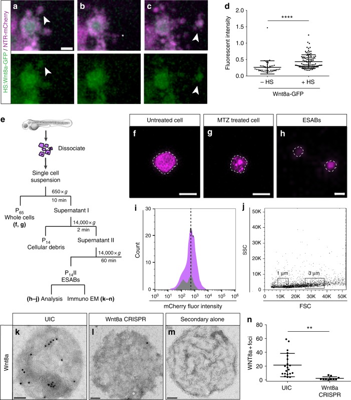
Apoptosis-induced stem cell division requires Wnt8a on the surface of ESABs (epithelial stem cell-derived apoptotic bodies). ac Time-lapse confocal images of an epithelial stem cell producing an apoptotic body containing Wnt8a-GFP (scale = 5 µm), see Supplementary Movie 4. d Quantitation of green fluorescent protein (GFP) fluorescence in ESABs after overexpression of Wnt8a by heat-shock induction. e Schematic of strategy to isolate ESABs by differential centrifugation. fh Whole cells (scale = 10 µm) and ESABs (scale = 5 µm) isolated by differential centrifugation exhibit mCherry fluorescence (magenta) and are appropriate size and morphology. i Quantification of the number of mCherry-positive ESABs after purification (magenta), compared to extracellular vesicles isolated from zebrafish larvae under homeostatic conditions (gray), using flow cytometry. j Size distribution of apoptotic bodies compared to size match bead controls (1 µm and 3 µm boxes, FSC = forward scatter, SSC = side scatter, see gating strategy in Supplementary Figure 6 a-b). km Transmission electron micrographs of Wnt8a immunogold labeling on whole-mount purified apoptotic bodies (scale = 500 nm). n The mean number of Wnt8a foci on individual ESABs from uninjected (n = 21) and Wnt8a CRISPR (clustered regularly interspaced short palindromic repeats)-injected (n = 10) animals. Data are from three independent experiments and error bars represent sd; ****p < 0.0001, **p < 0.001. Unpaired two-tailed t-test (d, n)

Acknowledgments
This image is the copyrighted work of the attributed author or publisher, and ZFIN has permission only to display this image to its users. Additional permissions should be obtained from the applicable author or publisher of the image. Full text @ Nat. Commun.