
|
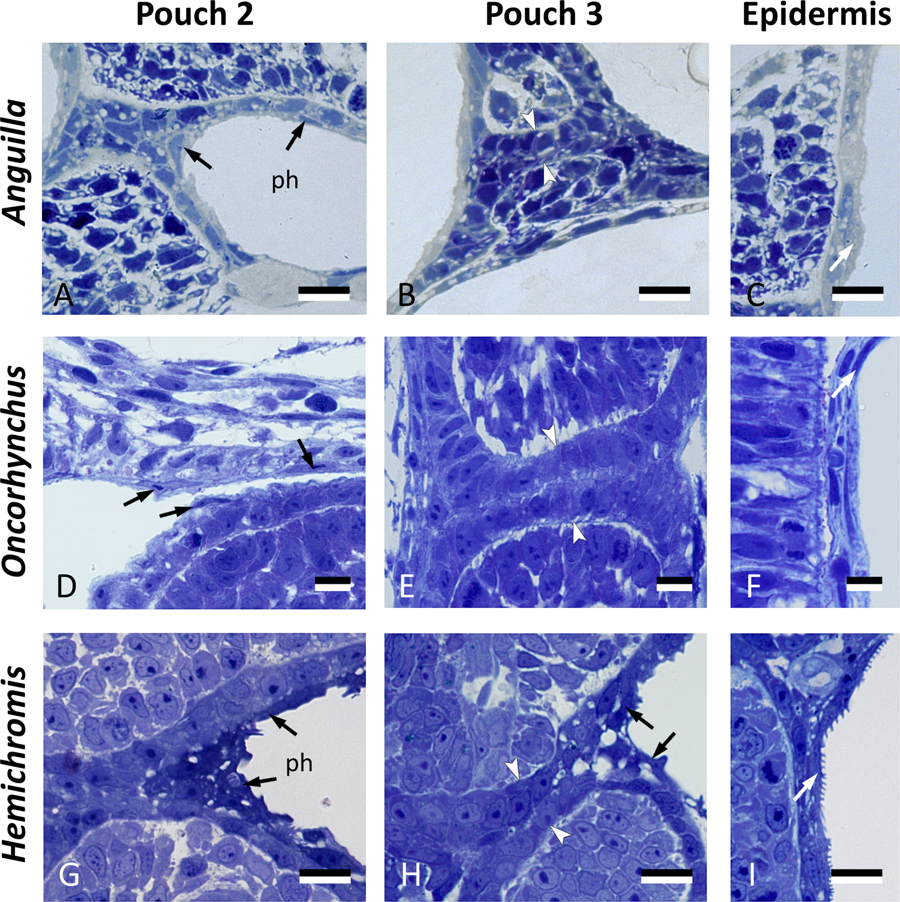
Fig. 6
Cells resembling periderm cells in the pharynx of other teleost species. Cross sections through the region of pouch 2, pouch 3 and comparison to the skin in three teleost species: Anguilla anguilla (total length, TL, 6.0 mm), Oncorhynchus tshawytscha (unhatched embryo, 15 dpf), and Hemichromis bimaculatus (TL 4.0 mm, 1 day post-hatching, dph). Pouch 2 (A,D,G) is lined by flattened cells (black arrows) overlying a layer of cuboidal cells. Where pouch 3 is not open yet (B,E), only a double layer of cuboidal endodermal cells is present (between white arrowheads). Where pouch 3 starts to open (H), similar cells can be seen to cover the pouch endoderm (black arrows). Note resemblance, for each of the three species, to peridermal cells in the superficial skin cover (white arrows, C,F,I). In (A,B,D,E,G,H), the epidermis is to the left and the pharyngeal lumen to the right; in (C,F,I) the external surface is to the right. (A–I) cross sections. ph: pharyngeal lumen. Scale bars (A–I) = 10 μm.