
|
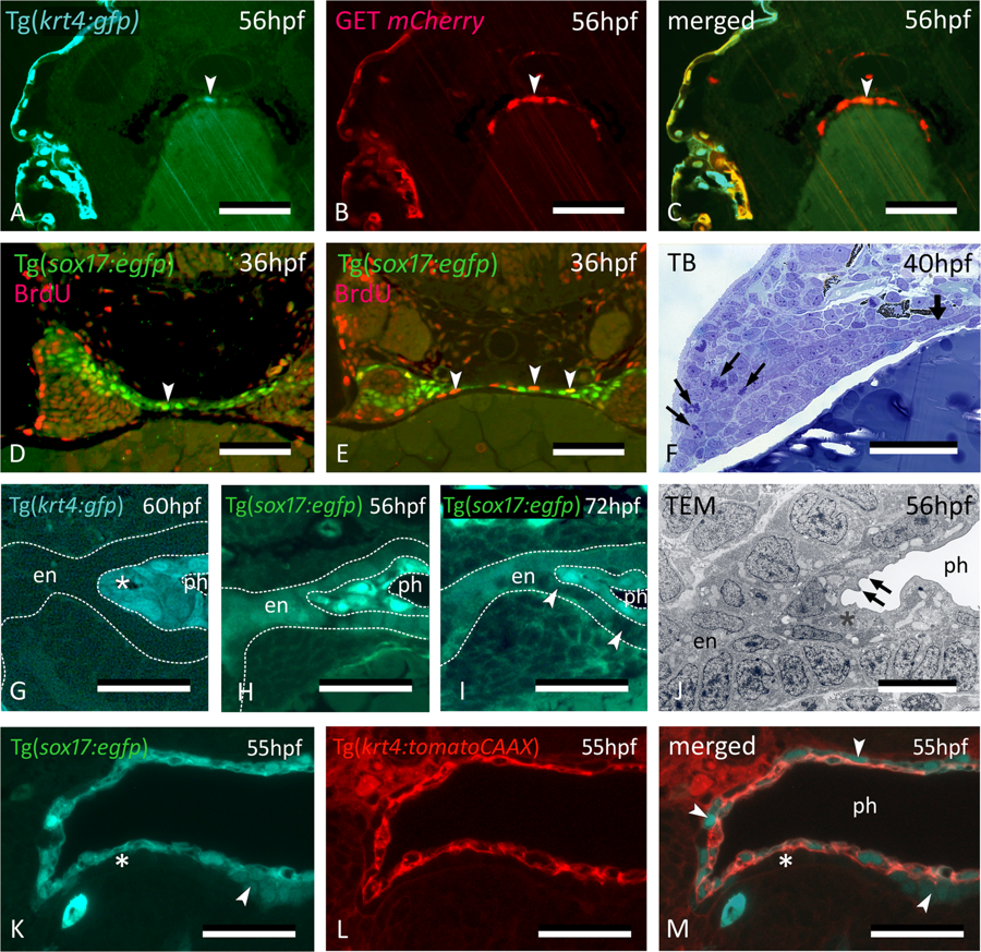
Fig. 4
Midline krt4+ cells are distinct from peridermal and endodermal cells. (A–C) In embryos of GET-periderm x Tg(krt4:gfp) line, at 56 hpf, both the periderm and the midline krt4+ cells (arrowhead) strongly express mCherry in patterns overlapping with the krt4:GFP + cells. (D,E) Mitoses (arrowheads) revealed by BrdU pulse labeling at 36 hpf in the midline endoderm at more anterior (D) and posterior (E) cross sectional level. (F) Abundant mitoses in the distal part of the pouch (arrows) but not in the midline endoderm (thick arrow). (G) Midline cells (star) maintain a strong krt4+ expression at 60 hpf. (H,I) Downregulation of sox17+ expression in the endodermal layer (delimited by the outer dotted line in H, and by arrowheads in I) but upregulation in the superficial layer surrounding the pharyngeal lumen (delimited by inner two dotted lines). (J) Cells of the superficial layer are flattened and more electron-dense in TEM than the basal endodermal layer, composed of cuboidal cells, and they develop microridges (arrows). Epidermis to the left; midline to the right. (K–M) Double transgenic embryo Tg(sox17:egfp;krt4:tomato) of 55 hpf; krt4+ cells are positive for sox17; conversely, only some cells of the basal endodermal layer have retained a sox17 signal (arrowheads). (A–M) cross sections. en: endodermal layer; ph: pharyngeal lumen; TB: toluidine blue staining. Scale bars (A–F) = 50 μm, (G–I) = 25 µm, (J–M) = 10 µm.