
|
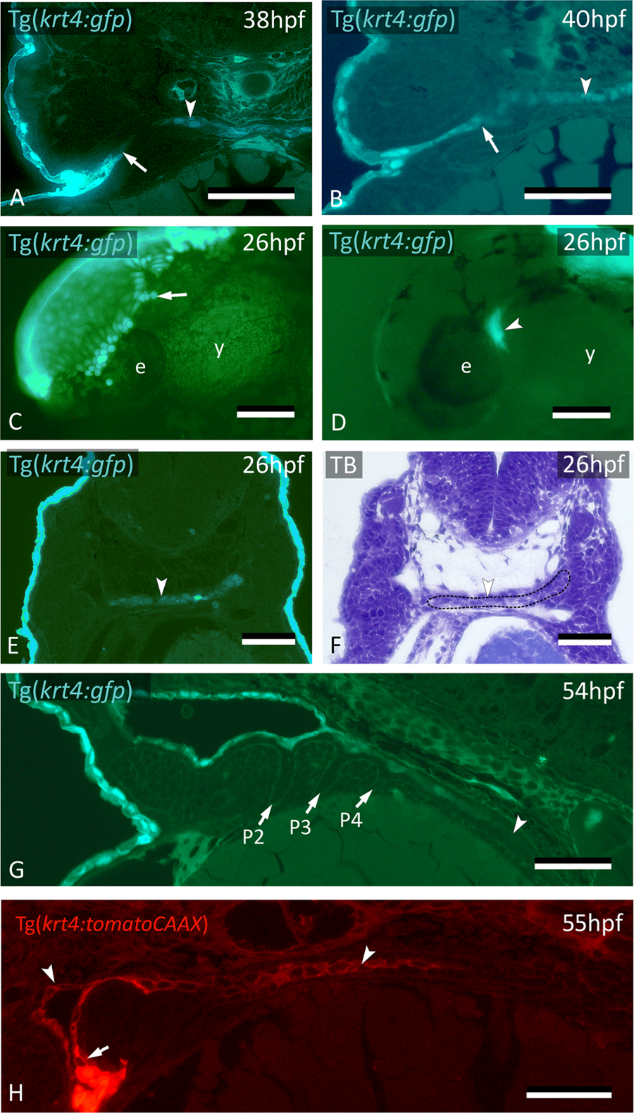
Fig. 3
krt4+ cells expand along the midline and connect to invaded peridermal cells. (A) At 38 hpf, krt4+ cells are visible along the midline (arrowhead), separated from invading peridermal cells (arrow). (B) Shortly after, the midline krt4+ cells (arrowhead) make contact with peridermal cells that have entered through P2 (arrow). (C) Lateral view on Tg(krt4:gfp) embryo in the early phase of periderm removal with EDTA. Part of the periderm is still attached (arrow). (D) At an advanced stage of EDTA treatment, the periderm is completely removed, and an internal krt4+ structure has become visible (arrowhead). (E,F) This internal krt4+ structure (arrowhead on E) coincides with the midline endoderm at the level of pouch 1–2 (arrowhead on F). (G) By 54 hpf, midline krt4+ cells cover the entire pharynx lining (boundary indicated by arrowhead). P2 is wide open (at another level of sectioning) but other pouches (P3–P6) are still free of krt4+ cells. (H) Double transgenic embryo Tg(sox17:egfp;krt4:tomato) of 55 hpf shows lining of pharyngeal epithelium with krt4+ cells (arrowheads) (red channel only). Note continuity, albeit with sharp boundary (arrow), with peridermal cells entering via P2. (A,B,E,F) Cross sections; (C,D) whole mount embryos; (G,H) sagittal section. TB: toluidine blue staining. e: eye; y: yolk; P2–P6: pouches 2 to 6. Scale bars (A,B,E–G) = 50 μm; (C,D) = 100 μm; (H) = 20 µm.