
|
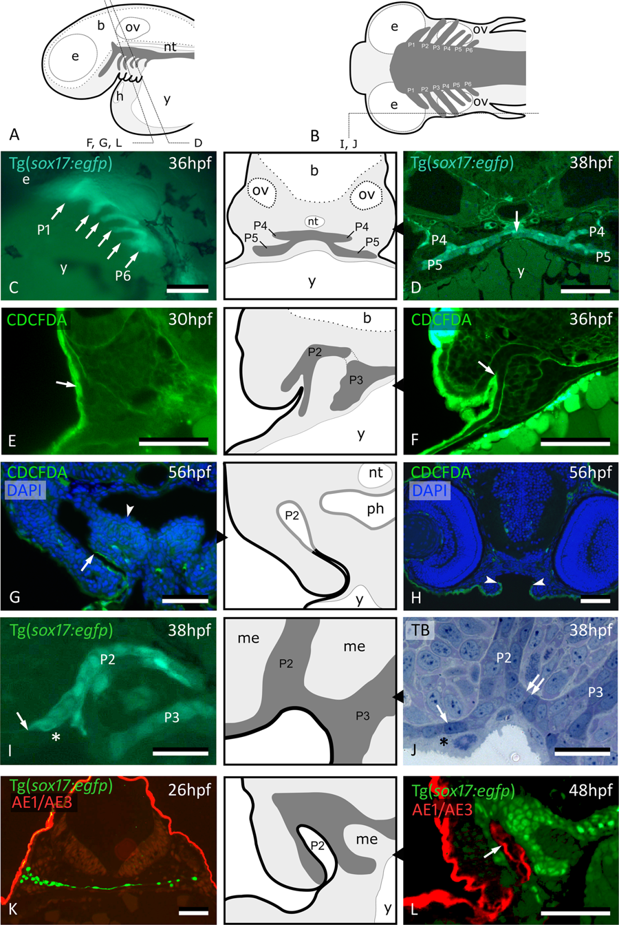
Fig. 1
Peridermal cells invade pouch 2. (A,B) Generalized scheme of zebrafish pouches (dark grey) in a lateral (A) and horizontal (B) view, with sections D-L as indicated. (C) Lateral view of Tg(sox17:egfp) embryo showing pouches 1 to 6 (compare to (A)). (D) GFP labeled endodermal cells along the midline (white arrow) extend into pouches 4 and 5 (P4 and P5, resp.); corresponding schematic attached. (E) CDCFDA labeling at 26 hpf, sacrifice at 30 hpf; labeled cells are exclusively located in the superficial skin layer or periderm (arrow, level of pouch 2). (F) CDCFDA labeling at 26 hpf, sacrifice at 36 hpf; peridermal cells have partially invaded pouch 2 (arrow); corresponding schematic attached. (G) CDCFDA labeling at 26 hpf, sacrifice at 56 hpf; peridermal cells (arrow) line the now open gill slit; elsewhere the pharyngeal epithelium remains free of peridermal cells (arrowhead); corresponding schematic attached. (H) CDCFDA labeled cells only cover part of the epithelial lining of the mouth. Their boundary is indicated by arrowheads and coincides with the reduction of two to one epithelial cell layer. (I,J) Contact area of pouches 2 and 3 with skin; note that sox17+ cells spread out (arrows) and appear to displace the basal cell layer. In this way, the distal parts of the pouches even become connected (double arrow). Only periderm (asterisks) covers the expansion of sox17+ endodermal cells. Corresponding schematic attached. (K,L) Immunolabeling using the pan-cytokeratin marker AE1/AE3 on Tg(sox17:egfp) embryos labels periderm only at 26 hpf (K), but also marks the lining of P2 at 48 hpf (arrow) (L); corresponding schematic attached. In all schemes, endoderm is dark gey, periderm is a black line. (C) Whole mount embryo; (D–H,K,L) cross sections; (I,J) sagittal sections. b: brain; e: eye; h: heart; me: mesenchyme; nt: notochord; ov: otic vesicle; P2 – P6: pouches 2 to 6; ph: pharyngeal lumen; TB: toluidine blue staining; y: yolk. Scale bar (C) = 500 μm, (D–H,K,L) = 50 μm, (I,J) = 20 µm.