
Image
|
Figure Caption
Fig. S4
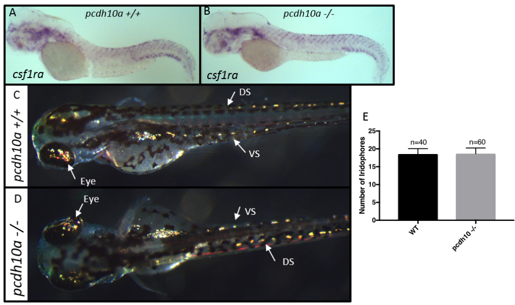
Removal of pcdh10a does not cause reduction of iridophores or xanthoblasts. (A, B) ISH at 2 dpf for xanthoblast marker csf1ra in (A) wildtype embryos, (B) pcdh10a -/- mutant embryos. (C,D) Incident light images of 3 dpf embryos to show yellow reflective iridophores in the eye, dorsal stripe (DS), and ventral stripe (VS). (E) Quantification of iridophores shows there was no significant difference in the number of iridophores located in the dorsal stripe.
Figure Data
Acknowledgments
This image is the copyrighted work of the attributed author or publisher, and
ZFIN has permission only to display this image to its users.
Additional permissions should be obtained from the applicable author or publisher of the image.
Reprinted from Developmental Biology, 444 Suppl 1, Williams, J.S., Hsu, J.Y., Rossi, C.C., Artinger, K.B., Requirement of zebrafish pcdh10a and pcdh10b in melanocyte precursor migration, S274-S286, Copyright (2018) with permission from Elsevier. Full text @ Dev. Biol.