
|
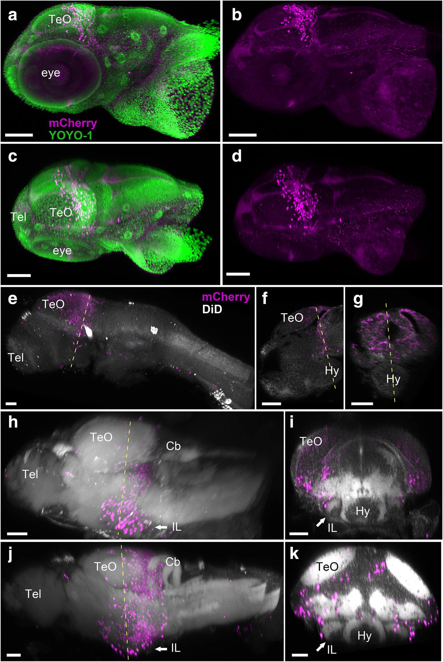
Fig. 4 Localization of the mCherry-positive cells in young larval brains of Tg(her5:ERT2CreERT2;βact:lox-stop-lox-hmgb1:mCherry) zebrafish treated with tamoxifen at 24 hpf. Anterior to the left for a–d, e, h, and j. a–d 3D reconstruction from confocal images of a whole head of 3 dpf larva. mCherry-positive cells are shown in magenta, and YOYO-1, a nuclear marker, is shown in green. a, b Side view of the larval head with (a) and without (b) YOYO-1 labeling. c, d Top view of the larval head with (c) and without (d) YOYO-1 labeling. The mCherry-positive cells are still close to the MHB at this stage. Some cells are starting to migrate anteriorly, but there are no mCherry-positive cells in the forebrain or in other brain areas. e–k 3D reconstruction from confocal images of dissected brains of 3 dpf (e–g), 5 dpf (h, i), and 7 dpf (j, k) larvae. mCherry-positive cells are shown in magenta, and DiD fiber labeling is shown in gray. e A whole brain at 3 dpf is shown in lateral view. f A sagittal section through the same specimen. g A frontal section. The hypothalamus (Hy) is extending in ventral position below the midbrain and is devoid of mCherry-positive cells. h A whole brain at 5 dpf is shown in lateral view. i A frontal section from the same brain showing the first appearance of the inferior lobe (IL; arrow), with a few mCherry-positive cells at the periphery of the structure. j A whole brain at 7 dpf is shown in lateral view. k A frontal section from the same brain showing the growing IL (arrow), with more mCherry-positive cells added laterally. Abbreviations: Cb cerebellum, Hy hypothalamus, IL inferior lobe, TeO optic tectum, Tel telencephalon. Scale bars: a–d, 100 μm. e–k, 50 μm