
|
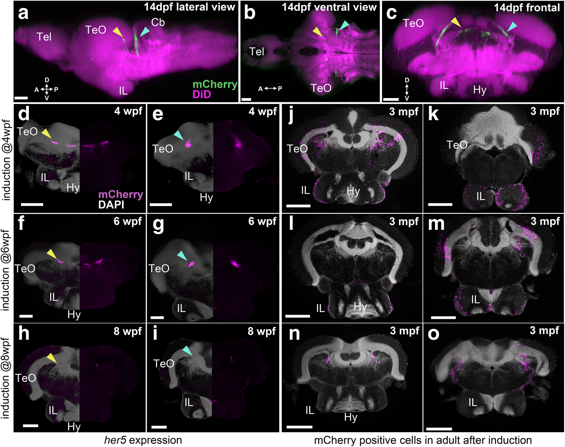
Fig. 8
Localization of her5 progenies following the induction at juvenile stages. a–i The brains of Tg(her5:mCherry) zebrafish to indicate the her5 expression at late larval to juvenile stages. j–o The brains of Tg(her5:ERT2CreERT2;βact:lox-stop-lox-hmgb1:mCherry) to indicate their progenies in the adult (3 mpf) brains. a–c 3D reconstruction from confocal images of a 14 dpf brain, showing mCherry (representing her5 expression) in green and DiD fiber labeling in magenta. Yellow arrows indicate the anterior her5-expressing domain, while blue arrows indicate the posterior her5-expressing domain. aThe whole brain in lateral view, b in ventral view, and c in a frontal section from the same 3D visualization. d–i Frontal sections of juvenile brains of Tg(her5:mCherry) (d, e at 4 wpf, f, g at 6 wpf, and h, i at 8 wpf), showing mCherry (representing her5 expression) in magenta and DAPI nuclear labeling in gray. The right half of the brain is demonstrated without DAPI to better visualize the mCherry signals. d, f, h The sections containing the anterior her5-expressing domain (yellow arrows). e, g, i The sections containing the posterior her5-expressing domain (blue arrows). j–o Frontal sections of 3 mpf brains of Tg(her5:ERT2CreERT2;βact:lox-stop-lox-hmgb1:mCherry), after tamoxifen induction at the corresponding juvenile stages (j, k are the brain induced at 4 wpf, l, m at 6 wpf, and n, o at 8 wpf). j, l, n The sections showing the anterior IL. k, m, o The posterior IL. Note that the mCherry labelings (magenta) in these sections represent progenies of the cells shown in d–i. Scale bar: a–c, 50 μm; d–g 100 μm; h, i 200 μm; j–o, 350 μm. Abbreviations: Cb cerebellum, Hy hypothalamus, IL inferior lobe, Tel telencephalon, TeO optic tectum