
|
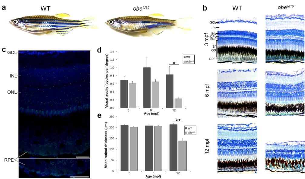
Fig. 1
Retinal structure and function in obetd15 zebrafish. (a) Wholemount morphology of adult wild-type (WT) and obetd15zebrafish. (b) Retinal histology of obetd15 zebrafish at 3, 6 and 12 months post fertilization (mpf). (c) Expression of kcnj13mRNA (green) in the WT adult zebrafish retina detected using an RNAscope assay. Sections are counterstained with DAPI nucleic acid stain (blue). (d) Visual acuity (cycles per degree) of obetd15 zebrafish at 3, 6 and 12 mpf, measured using optokinetic response assay (minimum n = 4, mean ± SEM). (e) Retinal thickness (µm) of obetd15 zebrafish at 3, 6 and 12 mpf, measured using OCT (n = 5 for each age, mean ± SEM). GCL, ganglion cell layer; IPL, inner plexiform layer; INL, inner nuclear layer; OPL, outer plexiform layer; ONL, outer nuclear layer; IS/OS, outer and inner segments; RPE, retinal pigment epithelium. *p < 0.05, **p < 0.01. Scale bars = 50 μm (b) and 25 μm (c).