
|
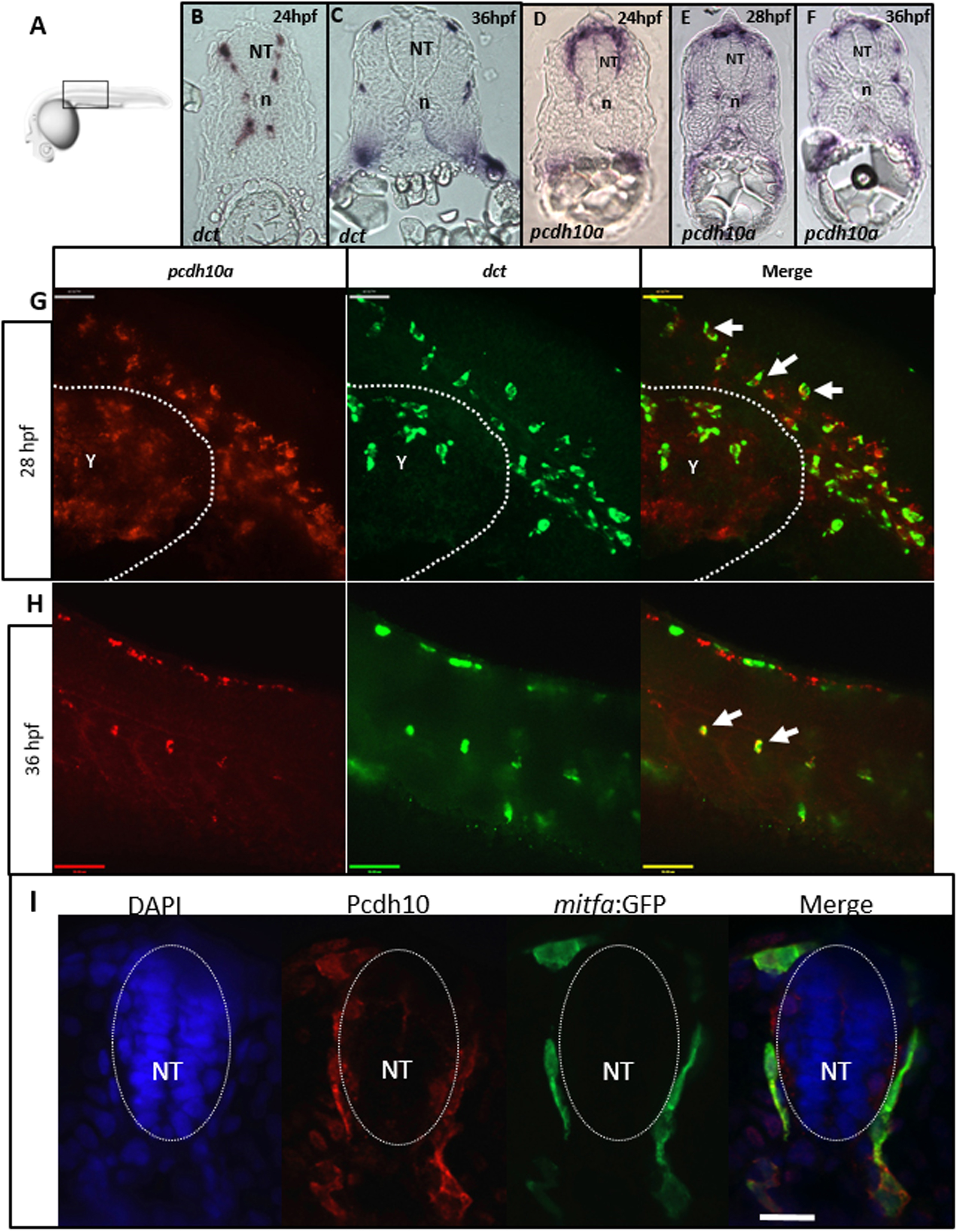
Fig. 2
pcdh10a is expressed in migratory NCC-derived melanocyte precursors. (A) Schematic for imaging region for B-I. (B-F) Cross-sectional analysis (12 µm) of ISH comparing the melanocyte marker dct with pcdh10a temporally represented across trunk NCC migration time points. (B, C) dct expression in wildtype embryos at 24 hpf and 36 hpf respectively. (D-F) pcdh10a expression in wildtype embryos at 24 hpf, 28 hpf, and 36 hpf respectively. The expression of pcdh10a mirrors that of dct during melanocyte precursor migration suggesting pcdh10a is expressed in melanoblasts. (G, H) Lateral view of whole embryo double FISH comparing expression and colocalization of pcdh10a (red) with dct (green). White arrows are examples of colocalization. Scale bars are 50 µm. Single Z-plane images. (I) 12 µm cross sections through the embryo trunk stained for Pcdh10 immunofluorescence. Scale bar is 20 µm, images are a single 1 µm Z-plane, scale bar is 20 µm. NT is neural tube, n is notochord. Anterior is to the left in G-H.
Reprinted from Developmental Biology, 444 Suppl 1, Williams, J.S., Hsu, J.Y., Rossi, C.C., Artinger, K.B., Requirement of zebrafish pcdh10a and pcdh10b in melanocyte precursor migration, S274-S286, Copyright (2018) with permission from Elsevier. Full text @ Dev. Biol.