
|
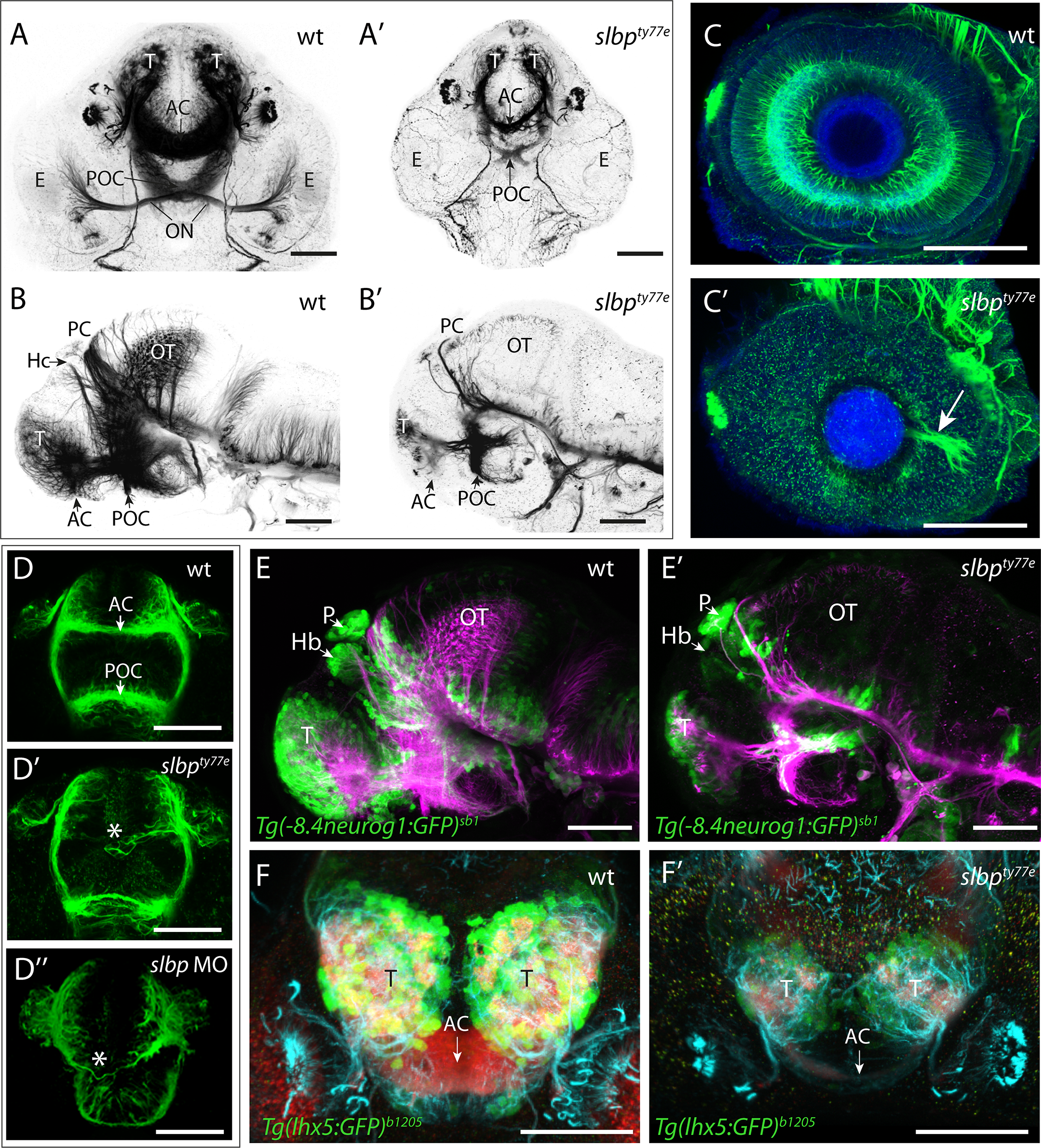
Fig. 2
(A-D”) Acetylated α-tubulin labelling of wildtype (A-D) and ele mutant (A’-D”) embryo brains and eyes. Frontal (A-A’; D-D”) and lateral (B- B’) views of brains and lateral views of eyes (C,C’) of 3dpf (A-C’) and 30hpf (D- D”) wildtype (A-D), slbpty77e mutant (A’-D’) and morphant (D”) embryos. Arrowhead in C’ highlights the aberrant extension of RGC axons within the retina. The asterisks in D’-D” highlight aberrantly positioned axons near the midline commissural region in slbp mutant and morphant embryos. (E-E’) Lateral view with anterior to the left of 3dpf wildtype (E) and slbpty77e mutant (E’) embryos showing expression of the Tg(-8.4neurog1:GFP)sb1 transgene (green) labelled neurons and acetylated α-acetylated tubulin labelled axons/neurites (magenta). (F-F’) Frontal views of the telencephalon in 3dpf wildtype (F) and slbpty77e mutant (F’) embryos showing expression of the Tg(lhx5:GFP)b1205 transgene (green) labelled neurons, SV2 labelled neuropil (red) and acetylated α-acetylated tubulin labelled axons/neurites (cyan). Abbreviations: AC, anterior commissure; POC, post-optic commissure; ON, optic nerve; T, telencephalon; PC, posterior commissure; OT, optic tectum; Hb,habenula; E,eye; P,pineal. Scale bars: (A-C”, E, E’) 100μm (D-D”, F, F’) 50μm