
Image
|
Figure Caption
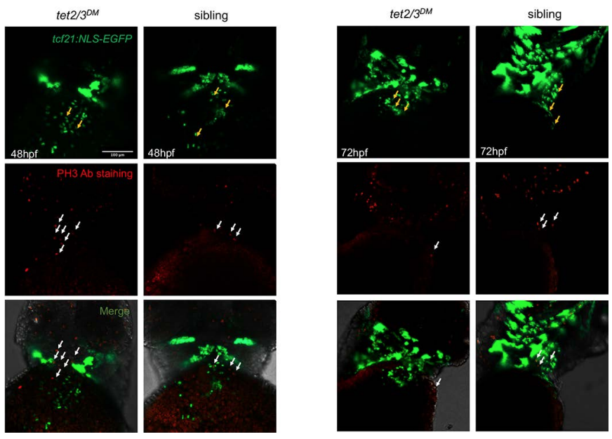
Fig. S3
No PE Cell Proliferation at 48 and 72 hpf Sibling and tet2/3DM Larvae. Related to Figure 1.Confocal images showing sibling and tet2/3DM larvae carrying the Tg(tcf21:NLS-EGFP) transgene stained by anti-GFP antibody (Green) plus anti-pH3 antibody (Red). Yellow arrows indicate PE cells. White arrows indicate pH3+ proliferating cells. Co-staining shows essentially no cell proliferation of tcf21+ PE cells at 48-hpf or 72-hpf.
Scale bar: 100 μm.
Acknowledgments
This image is the copyrighted work of the attributed author or publisher, and
ZFIN has permission only to display this image to its users.
Additional permissions should be obtained from the applicable author or publisher of the image.
Full text @ Cell Rep.