
|
Fig. 2
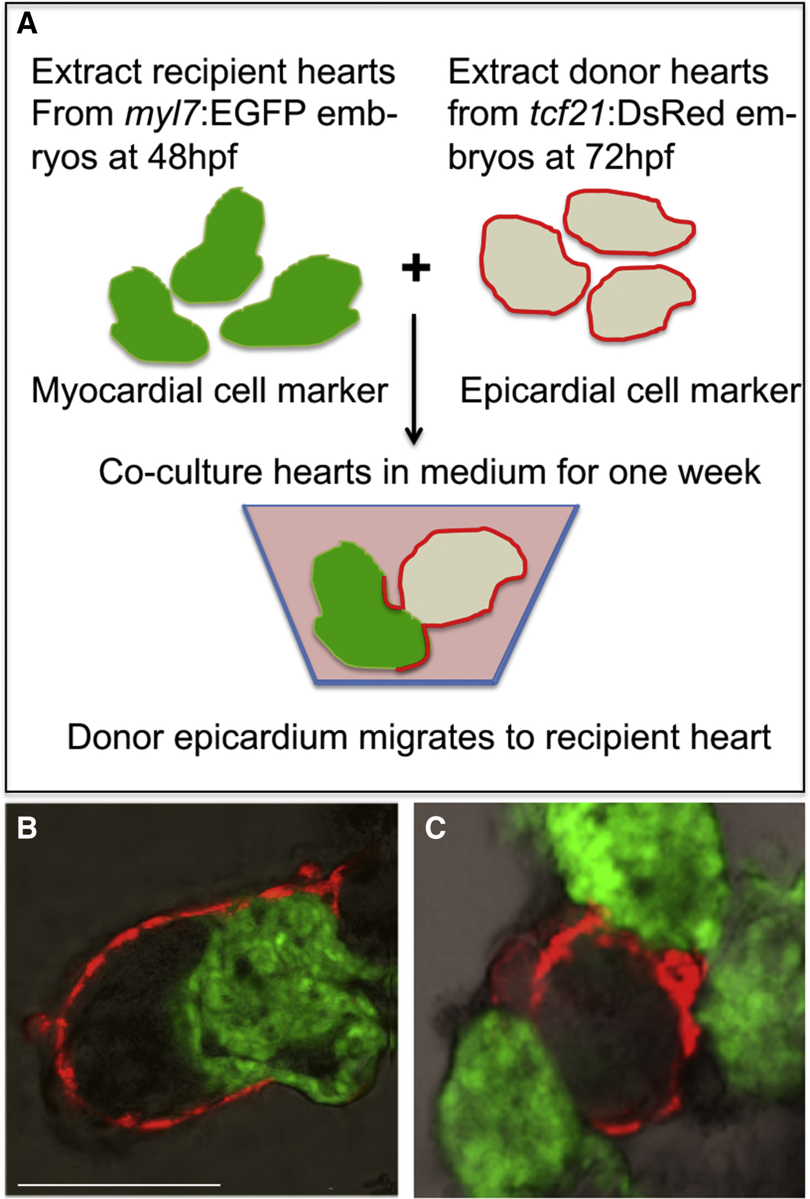
Epicardial Cells from Donor Hearts Do Not Migrate onto tet2/3DM Recipient Hearts
(A) Schematic of epicardial cell migration assay. Wild-typetcf21:DsRed donor hearts (isolated at 72 hpf) were co-cultured with either wild-type or tet2/3DM myl7:GFP recipient hearts (isolated at 48 hpf) for one week and then confocal images taken.
(B) Confocal images showed epicardial cells from wild-type donor heart can migrate onto wild-type recipient hearts. In 30 pairs of co-cultured hearts, 6 of recipient hearts were observed having epicardial cells migrated from donor hearts.
(C) Confocal images showed epicardial cells from wild-type donor heart failed to migrate onto tet2/3DM recipient hearts. In 60 pairs of co-cultured hearts, none of the recipient hearts were observed having epicardial cells from donor hearts.
Scale bar: 50 μm.