
|
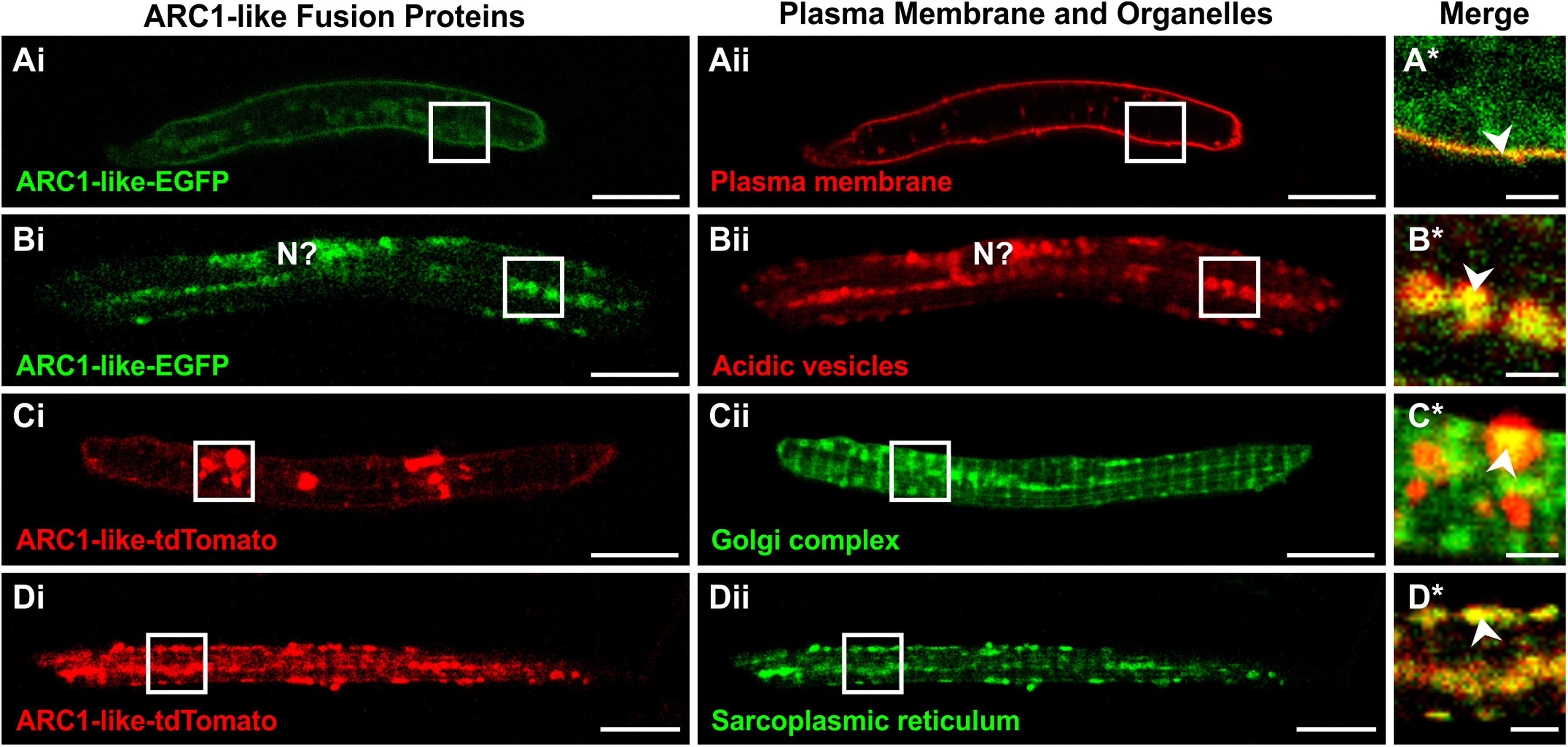
Fig. 3
Localization of ARC1-like fusion proteins in zebrafish primary cultured muscle cells. (A–D) Approximately 100–200 ng of either arc1-like-EGFP mRNAor arc1-like-tdTomato mRNA was injected into the blastodisc of zebrafish embryos at the one-cell stage to transiently overexpress the ARC1-like fusion proteins. At ~ 48 hpf, the embryos were digested and primary cultures were prepared from the dissociated cells. Primary cultured muscle cells that expressed (Ai, Bi) ARC1-like-EGFP were then labelled with (Aii) FM 6–46 (n = 24) or (Bii)LysoTracker Red DND-99 (n = 44) to visualize the plasma membrane or acidic vesicles, respectively, whereas those expressing (Ci, Di) ARC1-like-tdTomato were labelled with (Cii) BODIPY FL C5-ceramide (n = 16) or (Dii) ER-Tracker Green (n = 14) to visualize the Golgi complex or sarcoplasmic reticulum (SR), respectively. These are single optical sections to show the distribution of (Ai, Bi) ARC1-like-EGFP expression (in green) with respect to (Aii) the plasma membrane or (Bii) acidic vesicles (both in red), or the distribution of (Ci, Di) ARC1-like-tdTomato expression (in red) with respect to (Cii) the Golgi complex or (Dii) the sarcoplasmic reticulum (both in green). The regions bounded by the white squares in panels (Ai–Aii, Bi–Bii, Ci–Cii, Di–Dii)are shown at higher magnification and when the green and red channels are merged in the panels (A*–D*), respectively. In the merged images in panels (A*–D*), overlapping regions are shown in yellow. The arrowheads in panels (A*–D*)indicate overlap in fluorescence (yellow) between the ARC1-like fusion proteins and the various cellular compartments. “N?” in panel (Bi, Bii) is the putative location of the nucleus. Scale bars, 10 µm (in panels Ai–Aii, Bi–Bii, Ci–Cii, Di–Dii); and 2 µm (in panels A*–D*).
Reprinted from Developmental Biology, 445(2), Kelu, J.J., Webb, S.E., Galione, A., Miller, A.L., Characterization of ADP-ribosyl cyclase 1-like (ARC1-like) activity and NAADP signaling during slow muscle cell development in zebrafish embryos, 211-225, Copyright (2018) with permission from Elsevier. Full text @ Dev. Biol.