
|
Fig. 1
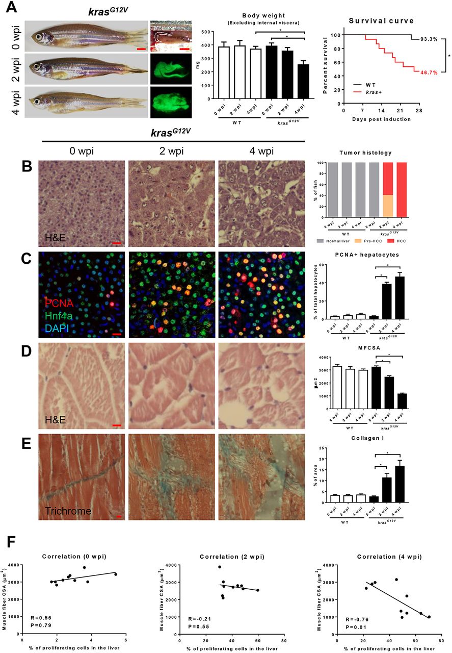
Characterization of zebrafish muscle wasting in krasG12V-induced HCC zebrafish.4-month-old male adult krasG12V and WT zebrafish were treated with dox for 4 weeks and sampled at 0 wpi, 2 wpi and 4 wpi. In each group, 15 fish were used to initiate the experiments. (A) Gross appearance and liver morphology (left), body weight excluding internal viscera (middle) and survival curves (right). (B) H&E staining of liver sections of krasG12V fish. Quantification of tumor histology (right). (C) IF staining of PCNA (red), Hnf4a (green) and DAPI (blue) in liver sections of krasG12V fish. Quantification of percentage of proliferating hepatocytes (right). (D) H&E staining of muscle sections of krasG12V fish. Quantification of MFCSA (right). (E) Gomori's trichrome staining of muscle sections of krasG12V fish. Quantification of percentage of collagen deposited area (right). (F) Correlation between percentage of proliferating cells in the liver (x-axis) and MFCSA (y-axis) at 0 wpi (left), 2 wpi (middle) and 4 wpi (right). *P<0.05. Scale bars: 2.5 mm in A; 10 μm in B-E.