
|
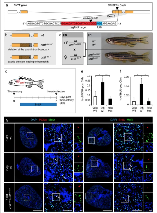
Fig. 6
Cntf mutants fail to enhance CM proliferation after thoracotomy. a Schematic drawing of the cntf locus containing 4 exons. UTR (white); translated sequences (orange boxes). The sequence of 154-174 nucleotides (in red) flanking PAM sequence (in blue) in the 3rd exon was targeted by the CRISPR/Cas9-sgRNA RNP complex. b Schematic drawing of the two deletions induced by CRISPR/Cas9. cntfdel207 contains a deletion of 207 bp in the 3rd exon spanning the exon/intron boundary. cntfdel7 comprises a deletion of 7 bp in the middle of the 3rd exon leading to a frameshift and a premature stop codon. c Genotypes and images of wild type and CNTF mutant siblings. F0 mosaic heterozygous candidates were crossed to obtain F1 trans-heterozygous mutants, which are viable without visible phenotype. Scale bar, 5 mm. dExperimental design. e Quantification of PCNA-positive cells among Mef2/DAPI-positive CM nuclei. n ≥ 3 hearts; ≥ 3 sections per heart; *P < 0.05, **P < 0.01. f Quantification of BrdU-positive cells among Mef2/DAPI-positive CM nuclei. n ≥ 3 hearts; ≥ 3 sections per heart; *P < 0.05. g, h Transversal heart sections of wt and cntf mutant fish at 7 dpt, immunostained against Mef2 (green, a myocyte nuclear marker) and cell proliferation markers, PCNA (g, red) or BrdU (h, red). All nuclei are labeled with DAPI (blue). Arrows indicate some proliferating CMs. Scale bar for the whole section, 500 μm; for the magnified area, 100 μm; for the zoom of magnified area, 20 μm