
|
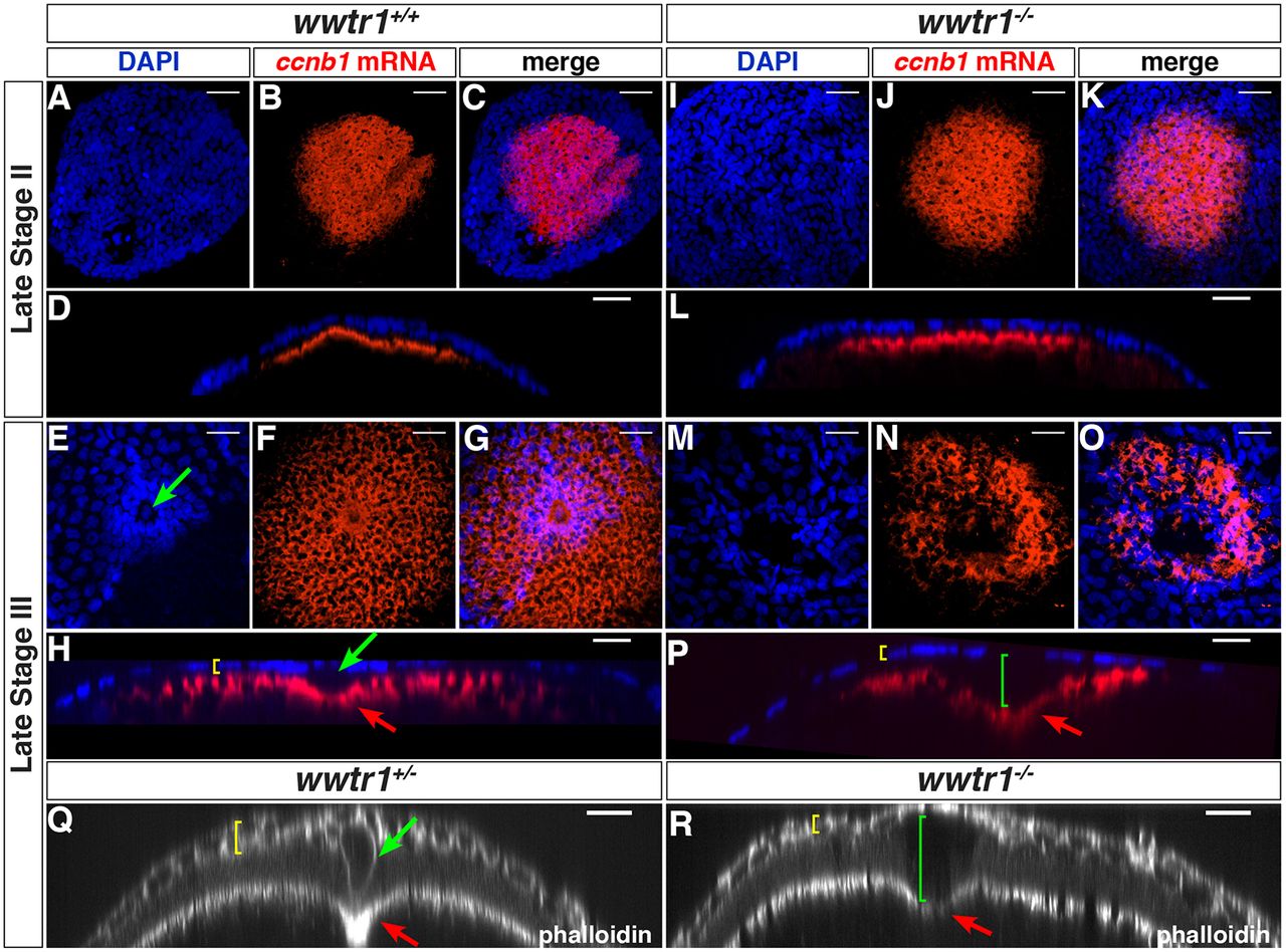
Fig. 3
wwtr1 mutant follicles lack the micropylar cell. (A-P) Maximum intensity projection of confocal images of wwtr1+/+ (A-H, n=31) or wwtr1−/− (I-P, n=40) follicles stained by ISH to detect ccnb1 mRNA at the animal pole (red), combined with DAPI nuclear staining (blue) at the indicated oocyte stages. (D,L,H,P) Orthogonal views of corresponding confocal stacks (C,K,G,O). (Q-R) Orthogonal views of confocal stacks of wwtr1+/− (Q, n=9) or wwtr1−/− (R, n=7) follicles stained with Rhodamine Phalloidin. Green arrows indicate an MC; red arrows indicate the indentation of the oolemma; yellow brackets indicate the FCL; green brackets indicate the gap between the FCL and the oocyte surface in wwtr1−/− follicles. The zp0.5:egfp-zorba transgene (not shown) was used to localize the animal pole and orient the follicles. Scale bars: 20 μm.